
Cultivation
Anaerobic cultivation techniques were applied18. One liter of freshwater medium contained 1.0 g of NaCl, 0.4 g of MgCl2 × 6 H2O, 0.1 g of CaCl2, 0.5 g of KCl, 0.2 g of KH2PO4, 0.25 g of NH4Cl, and 0.2 g of NaSO4 in pure water. After autoclaving, 1 ml of the following sterile solutions was added to 1 l of medium: chelated trace element mixture (per liter of distilled water: 2100 mg of FeSO4 × 7 H2O, 60 mg of H3BO3, 1.0 g of MnCl2 × 4 H2O, 0.38 g of CoCl2 × 6 H2O, 0.24 g of NiCl2 × 6 H2O, 2 mg of CuCl2 × 2 H2O, 0.29 g of ZnSO4 × 7 H2O, 72 mg of NaMoO4 × 7 H2O, and 7.8 g of Na2EDTA, pH 6.0), selenite-tungstate solution (per liter: 0.4 g of NaOH, 32 mg of Na2WO4 × 2 H2O, 24 mg of Na2MoO4 × 2 H2O, and 6 mg of Na2SeO3 × 5 H2O), vitamin solution (4 mg of 4-aminobenzoic acid, 2 mg of D-(+)-biotin, 10 mg of nicotinic acid, 5 mg of calcium D-(+)-pantothenate, 15 mg of pyridoxin hydrochloride, 4 mg of folic acid, and 1.5 mg of lipoic acid in 100 ml of 10 mM NaH2PO4, pH 7.1), cyanocobalamin solution (5 mg L− 1), thiamine solution (10 mg of thiamine hydrochloride in 100 ml of 25 mM NaH2PO4, pH 3.4), and riboflavin solution (2.5 mg in 100 ml of 25 mM NaH2PO4, pH 3.2). After addition of sterile stock solutions − 2 mL of 0.5 M cysteine, 2 mL of 1 M sodium acetate and 30 mL of 1 M NaHCO3 – the pH was adjusted to 7.0 with sterile 2.0 M HCl.
The methanogenic enrichment culture was maintained with an annual transfer of 10% (vol/vol) inoculum. The cultures contained 300 mL freshwater medium, 0.3 mL of 200 mM FeS suspension, 30 mL 2,2,4,6,8,8-heptamethylnonane (HMN), and 1.5 mL of R-(+)-limonene under an oxygen-free N2–CO2 atmosphere (90:10, vol/vol, < 7 ppm O2) in 500-mL borosilicate bottles. Incubation occurred on an orbiting shaker with 60 rpm at 28 °C5.
Cell separation and RNA extraction
Biomass was obtained by differential centrifugation. Large and aggregated cells were pelleted in a SW28 ultracentrifuge rotor (Beckman, Palo Alto, CA) at 7600 rpm (7643 g) for 20 min (10,000 Svedberg [S]). The 10 kS cell pellet was resuspended in 1 mL of 10 mM Tris, 1 mM EDTA, pH 8.0 (TE). RNA was extracted using the RNA PowerSoil Total RNA Isolation Kit (Qiagen, Hilden, Germany). RNA sequencing was performed by the Max Planck-Genome-centre Cologne, Germany (https://mpgc.mpipz.mpg.de/home/) using NEB Next Ultra RNA kit (New England Biolabs, Ipswich, MA) for the production of an Illumina-compatible library and Illumina HiSeq (Illumina, San Diego, CA) (150 bp reads; 57,878,109 reads revealing 8,918,223,863 bases were deposited at NCBI SRR30230645)6.
In situ hybridization
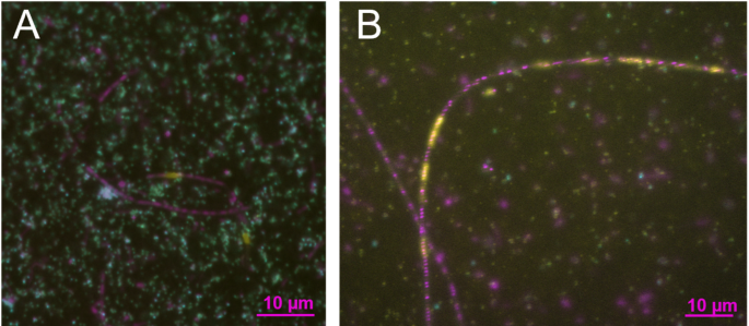
Catalyzed reporter deposition-fluorescence in situ hybridization (CARD-FISH)19 was used targeting 16 S ribosomal RNA and intron RNA. Living Methanosaeta cells were identified with probe ARCH915 (GTGCTCCCCCGCCAATTCCT)20. Ca. Velamenicoccus archaeovorus probe OP3-565 (TACCTGCCCTTTACACCC)21 was used together with manually designed partially degenerated helper oligonucleotides H548-A (AATAAATCCGAGTAACGC), H548-C (AATCAATCCGAGTAACGC), H583-TC (CTCCCCACTTGTCAGGCCGCC) and H583-CT (CCTCCCACTTGTCAGGCCGCC)5. The group I intron in the 23 S rRNA was targeted within the gene for homing endonuclease. Probes and flanking helper oligonucleotides were designed using Primer3 v. 4.0.0 (http://sourceforge.net/projects/primer3/). Specificity was tested against the metagenome datasets of the enrichment culture6. Probes were hen1-2235 (CCGCCAAGTAGTAGCCGATT), hen2-2309 (AACTTTCCACGGTGACTTGT), hen3-2538 (TCTTTATCATTTCTGCCAGTTCG) and the reverse complement hen2-rc2309 (ACAAGTCACCGTGGAAAGTT). Helper were Hhen1-2210 (TCTGGTTTCATAAAGACCTCCTTCT), Hhen1-2255 (TCCTTCTCCGTCGGCAAAAC), Hhen2-2288 (AGTCTTGCCGTTGTCTGAAGG), Hhen2-2329 (CTGGGAGATGTTGAAACAAAGAGA), Hhen3-2516 (CAGAATTTCGCAAAATCCCGTT), Hhen3-2561 (GCTACTCTTAAGATGTTCCCCCT). For intron detection, a formamide concentration of 20% was used at concentrations of 0.17 ng/µl for each probe and helper. One mL of culture was fixed with formaldehyde (1.3% [wt/vol]) for 60 min at 21 °C. Five µL of fixed culture were dried on a glass slide and then rinsed with 1 ml of phosphate buffered saline (PBS, pH 7.4). Cells were embedded in 0.1% [wt/vol] low-melting agarose and air dried. Permeabilization was performed for 60 min at 37 °C by lysozyme (10 mg/mL) and proteinase K (200 µg/ml) for 2 min at 21 °C. Washing steps were performed as described19. Endogenous peroxidases were inactivated with 0.1 M HCl for 1 min at 21 °C, a washing with 1x PBS, and 3% H2O2 for 10 min at 21 °C. The probes were hybridized for 160 min at 46 °C, followed by incubation with Alexa448 or Alexa594-tyramide conjugates at 46 °C for 45 min in the dark. After DNA staining with 4′,6-diamidino-2-phenylindole (DAPI, 1 µg/mL), cell preparations were embedded in Citifluor-Vectashield antifading medium (4:1 [vol/vol]). Images were obtained with an epifluorescence microscope (Zeiss Axiophot with camera MRm and HBO100 light source using Zeiss Axiovision). Confocal laser scanning microscopy (CLSM) and super resolution structured illumination microscopy (SR-SIM) were performed using a Zeiss LSM 780 equipped with a ELYRA PS.1 module. Zeiss Zen 3.11 was used to analyse the images (Zeiss, Oberkochen, Germany).
Sequence analysis
RNA reads (NCBI: SRR30230645) were trimmed with BBDuk to > Q30 and longer than 30 nt. Coverage of reads mapping with at least 99% identity was determined using BBMap and Ca. Velamenicoccus archaeovorus (NCBI CP019384) or partial sequences thereof (300 nt for exon/intron border, intron sequence). Both calculations were performed within Geneious (Dotmatics, Boston, MA) with the default parameters within Geneious, except for the ones stated. Reads covering the borders between intron and exon were quantified by manual inspection of mapped and aligned reads (150 bp) to aforementioned partial sequences.