

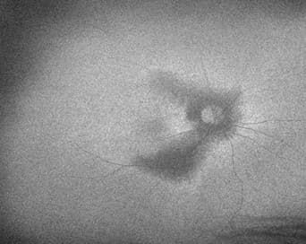
 Fig 2a–d. Ultra-widefield imaging (top two images) showing perivascular pigmentary changes and fundus auto-fluorescence (bottom two images) of the patient at presentation showing hypofluorescence due to RPE atrophy
Discussion
Pigmented paravenous retinochoroidal atrophy is a rare condition characterised by slowly progressive atrophy of the retinal pigmented epithelium (RPE) and the choriocapillaris. These changes are bilateral, often asymmetric and characterised by bone corpuscle pigment accumulation limited to the retinal vascular arcades and the peripapillary region1.
Although the exact mechanism remains unknown, many dysgenetic, degenerative, hereditary and inflammatory conditions have been associated with the disease. PPRCA was initially described in 1937 by Dr Hewitson-Brown as retino-choroiditis radiata in a patient with tuberculosis2. Further inflammatory associations include sarcoidosis, Behçet disease, measles, rubeola, syphilis and tuberculosis3.
As the majority of the patients are asymptomatic – uncommonly, patients may present with blurry vision in cases where the atrophy extends to include the foveal region4 – so initial diagnosis is based on incidental retinal findings during routine eye examinations.