
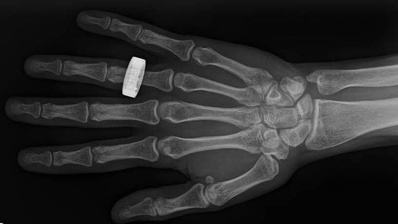
A new study reports that astronauts on a private SpaceX flight took the first diagnostic X-rays in space, capturing clear pictures of a hand, a chest, and other body parts while circling more than 200 miles above Earth.
None of the crew were radiologists, and they learned the technique in about four hours.

The result opens a door that stayed shut through six decades of human spaceflight. Crews bound for the Moon or Mars could soon spot a broken bone, a collapsed lung or worsening bone loss on their own.
They would no longer need to wait until they fly home, far from any hospital.
First X-rays taken in space
The images came from Fram2, a privately funded mission that in spring 2025 became the first to carry people in a polar orbit, looping over both of Earth’s poles. Its four-person crew spent about three days inside a SpaceX Dragon capsule.
Packing an X-ray suite into that cabin meant stripping the hardware down. The generator and detector together weighed about 25 pounds (11 kilograms). It ran on a rechargeable battery, so the crew needed no fixed power source or shielded room.
Doctors have wanted this capability for decades. Bringing X-rays to orbit has become its own small field of study. One recent review lays out why the tool suits the medical problems astronauts actually face.
The detector did something a standard X-ray machine cannot. From a single exposure, it produced separate views of bone and soft tissue. It did so at a radiation dose below one-thousandth of what an astronaut is cleared to absorb over a career.
For their first attempt, the crew X-rayed a hand wearing a ring. It was a deliberate echo of the first X-ray ever taken – made by Wilhelm Roentgen of his wife’s hand in 1895. The nod to history was intentional.
Portable X-ray fit inside the capsule
Over the following days they imaged the forearm, chest, pelvis and abdomen. They even X-rayed onboard electronics to see whether the same tool could check hardware for hidden faults.
The work was part of SpaceXray, a project run by physicians and engineers across North America. Its principal investigator is Dr. Sheyna Gifford, a physician at Saint Louis University (SLU).
She spent years writing the protocols crews would need. Her goal was a training plan simple enough for a non-expert to follow.
Overseeing the medical side was Dr. Lonnie Petersen, an associate professor at the Massachusetts Institute of Technology (MIT) who studies how spaceflight changes the body.
Petersen helped design a plan that could be taught quickly and carried out with almost no supervision.
Crew learned X-rays in four hours
The most striking part of the mission was how little preparation the imaging took. Radiographers on Earth train for years, yet the Fram2 crew were ready after a single afternoon of instruction.
The team used a train-the-trainer approach. One crew member learned the steps, taught the others, and then they photographed one another, checked each image on a screen and saved the good ones.
“The crew learned how to take X-rays in one afternoon,” said Petersen. Because the protocol had been written in advance, the astronauts could focus on aiming the machine rather than reading anatomy.
Quality was the real test. The images came back clear enough to read, with no repeat exposures needed, and by early assessments their sharpness held up well against what the same machine produced on the ground.
Researchers had long assumed that capturing a usable X-ray in orbit would demand a specialist’s skill and a steady setup.
Until this flight, no one had shown that ordinary crew members could do it in a cramped capsule floating in weightlessness.
Why astronauts need X-rays
Bone is the reason many researchers wanted X-rays in space at all. In microgravity the skeleton stops carrying weight, and astronauts can shed 1–2% of the bone density in their hip and spine each month.
That loss starts fast. A recent study found measurable changes in bone structure after flights lasting only a few days, the same short stretch the crew spent in orbit.
Ultrasound has been the only medical imaging tool aboard the International Space Station, and it shows bone poorly. That leaves a blind spot for exactly the injuries and changes crews are most likely to face.
A review of imaging options for spaceflight has argued that X-rays would fill that gap, catching fractures and early signs of thinning bone that ultrasound misses. The Fram2 detector goes a step further.
Because it separates bone from soft tissue in each exposure, it can measure bone density rather than just show it. That gives flight surgeons a number to track across a long mission, not merely a picture to eyeball.
What’s next for space medicine
The flight settled a question that had lingered since the 1960s. Diagnostic X-ray imaging works in orbit, and a lightly trained crew can produce pictures clear enough to guide real medical decisions.
The same hardware could watch over the spacecraft, not just the crew. On Earth, X-rays already check aircraft parts for hidden cracks. The crew’s test images of onboard electronics hint at a future where repairs in flight are guided by what the beam shows.
“Life in space will be safer and better because of this technology,” said Gifford, the project’s principal investigator.
The same portable tools could bring imaging to disaster zones, rural clinics, and other places on Earth where a full X-ray suite has not fit.
NASA is already testing compact X-ray systems for a possible flight to the International Space Station later this decade. What began as a hand and a ring in a passing capsule now looks like standard equipment for the long journeys ahead.
The study is published in Radiology.
Image credit: Radiological Society of North America
—–
Like what you read? Subscribe to our newsletter for engaging articles, exclusive content, and the latest updates.
Check us out on EarthSnap, a free app brought to you by Eric Ralls and Earth.com.
—–