
 Image credit: Steve Zill/Columbia Engineering
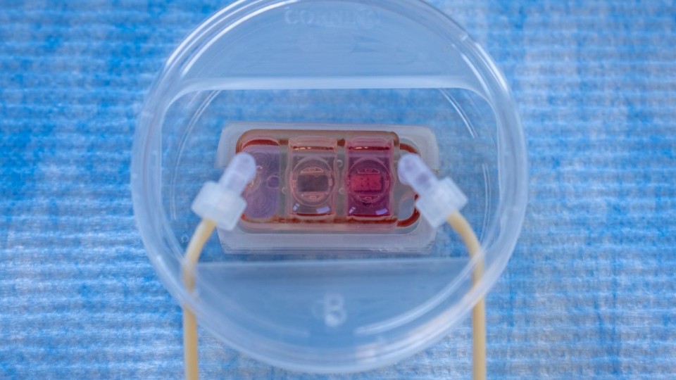
Researchers at Columbia University have developed a new multi-organ chip that recreates how cancer cells travel through the bloodstream and spread to distant organs
Metastasis occurs when cancer cells leave an original tumour, enter the bloodstream and establish themselves in another part of the body. It is responsible for at least two-thirds of cancer-related deaths.
The study, published in Science Translational Medicine, was led by Columbia Engineering professor Gordana Vunjak-Novakovic and a team of researchers from Columbia Engineering and the Herbert Irving Comprehensive Cancer Centre.
Bringing human tissues together
These tissues are connected through vascular circulation, allowing cancer cells to move between them in a way that resembles the human body.
The researchers engineered the bone, lung and blood-vessel lining from induced pluripotent stem cells. The tissues were grown and maintained using specialised scaffold and bioreactor systems designed to support their development and function.
A selectively permeable endothelial barrier separates the vascular channel from the tissue compartments. This barrier is particularly important because it recreates a major challenge cancer cells face when leaving the bloodstream and entering distant organs.
Tracking breast cancer cells
To test the system, the researchers introduced human breast cancer cells into the vascular circulation and observed how they interacted with the engineered organs.
The results showed that different cancer cells displayed distinct patterns of organ colonisation. Cells associated with bone metastasis showed stronger colonisation of the engineered bone and caused more pronounced bone degeneration.
Meanwhile, cells associated with lung metastasis caused greater disruption in lung tissue while showing comparatively limited colonisation of bone.
These differences suggest the multi-organ chip can reproduce key features of organ-specific metastasis, including certain cancer cells’ preference for particular tissues.
Preparing organs for cancer
The researchers also observed evidence of a process known as pre-metastatic niche formation. Before cancer cells establish themselves in a distant organ, they can alter the surrounding tissue and make it more favourable for future colonisation.
The engineered bone and lung tissues showed signs of conditioning by circulating cancer cells, allowing researchers to investigate how distant organs prepare for metastatic growth.
Animal models have been essential to cancer research, but differences between human and animal biology can make it difficult to predict how treatments will perform in patients.
The new multi-organ chip offers a complementary approach by using engineered human tissues that can be adapted to individual patients.
The researchers hope that their study will help uncover the molecular mechanisms behind metastasis and identify new therapeutic targets. The multi-organ chip could also contribute to the growing use of human-based laboratory models in preclinical research.