

A new pilot study suggests that tear film lipid irregularity, rather than tear volume alone, may be a key feature of dry eye disease associated with Sjögren’s syndrome (SS). The research used recently developed technology called the Tear Film Imager (TFI), a high-resolution, non-contact device that is not FDA-approved in the United States. The TFI uses spectral interference and reflective analysis of ultra-thin tear film layers to measure tear film dynamics while patients blink naturally. In this study, it showed that patients with Sjögren’s syndrome had significantly greater lipid map uniformity abnormalities than healthy controls, suggesting lipid layer dysfunction could be an important therapeutic target.
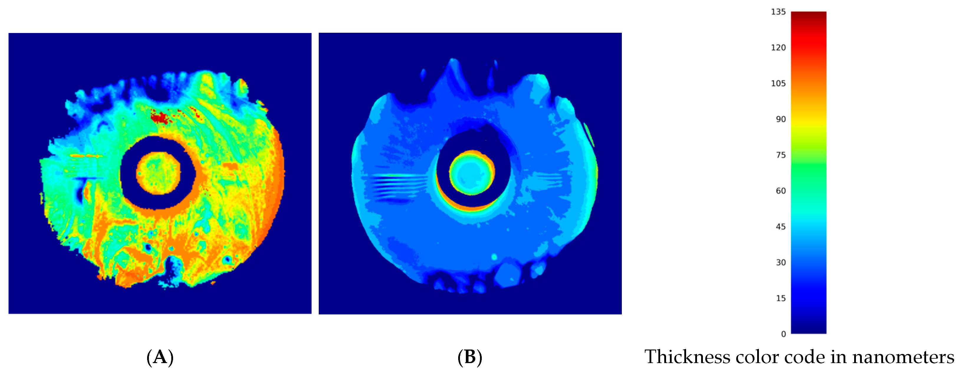
By capturing tear film sublayers dynamically and simultaneously, an investigational device called the Tear Film Imager revealed that lipid layer dysfunction may play a bigger role in the pathophysiology of DED in Sjögren’s patients than previously thought. These images from the study show a comparison of the lipid layer thickness maps between a participant with Sjögren’s syndrome (A) and a matched control subject (B). Photo: Goulak AM, et al. J Clin Med. 2026;15(18):7077. Click image to enlarge.
Published recently in Journal of Clinical Medicine, the cross-sectional study enrolled 15 patients with confirmed Sjögren’s syndrome and symptomatic dry eye disease (mean age: 53.4; 86.7% female) and compared them with 15 age-matched healthy controls (mean age: 52.3; 73.3% female). In addition to TFI imaging, Sjögren’s patients underwent Ocular Surface Disease Index testing, Schirmer’s testing with anesthesia, tear osmolarity measurement and tear breakup time. The controls underwent TFI imaging and anterior segment examination.
The TFI is designed to provide nanometer-resolution assessment of several characteristics of individual tear film layers: mucoaqueous layer thickness, muc-aqueous layer thinning rate, lipid layer thickness, lipid map uniformity, lipid breakup time and interblink interval during natural blinking. It also generates a 3D map of the lipid layer, allowing clinicians to visualize lipid uniformity across the tear film.
The main finding of this study was that lipid map uniformity was significantly higher in the Sjögren’s group than in controls (136.64nm² vs. 62.70nm², respectively), reflecting a more heterogeneous lipid distribution. The researchers explained in their paper that this “may suggest the presence of meibomian gland dysfunction amongst those with Sjögren’s Syndrome.” Moreover, these patients had a higher prevalence of lipid-related dry eye disease, “underscoring the role the lipid layer plays in the pathophysiology of dry eye in this population.”
In contrast, other mean TFI measurements were not significantly different between groups: muco-aqueous layer thickness was 3701.39nm in the Sjögren’s group vs. 3384.80nm in controls; lipid layer thickness was 72.08nm vs. 54.31nm; interblink interval was 11.63s vs. 7.33s; and lipid breakup time was 5.36s vs. 3.75s. These findings, the authors wrote, “further support the notion that lipid layer dysfunction may be more prominent in Sjögren’s Syndrome-related dry eye and that the lipid component of the tear film should be the focus of future research and therapeutic interventions” for the condition.
Moreover, the authors noted that tear film abnormalities captured by the TFI correlated with clinical findings, demonstrating the device’s potential “as a diagnostic and monitoring tool for assessing tear film function.” The team observed a strong positive correlation between Schirmer’s score and muco-aqueous layer thinning, “suggesting a connection between aqueous production and tear film thickness,” which they say “corroborates previous research utilizing the TFI.”
That said, the findings should be interpreted with caution. The Tear Film Imager is an investigational tool, and its clinical role remains to be established. This was also a small, single-center pilot study with only 15 patients in each group, so the results should be considered preliminary. Larger studies will be needed to confirm whether the device can reliably inform routine clinical care and whether the observed lipid-layer abnormalities translate into meaningful changes in diagnosis or treatment.
Overall, this study reinforces that Sjögren’s dry eye is not just about aqueous deficiency. Lipid layer instability may be a major contributor, so meibomian gland evaluation and lipid-targeted therapies may deserve greater attention. The authors argued that future studies “can continue this research by expanding the patient population and categorizing patients based on their systemic disease and ocular findings, further advancing our understanding of SS-related DED and its management.”
Click here for the journal source.
This article was developed by the editorial staff in conjunction with experts in the field. In the process, AI may have been among the editorial tools used to meet the goals of human editors, who approved all content.