
The 2026 edition of our AMA: Energy online conference returns on April 30th. Register now!
The Academy of Medical Sciences has awarded Dr. Robert Owen, a researcher at the University of Nottingham’s School of Pharmacy, a grant to study how physical surface geometry influences cell behavior, with the goal of building those cues directly into healthcare materials.
The grant is part of a £6.7 million tranche the Academy distributed across 55 early career researchers at 38 UK institutions through its Springboard program.
Owen’s project uses ultra-high-resolution 3D printing to build materials with precisely engineered surface features, such as microscale curves, and then observe how cells involved in bone healing and skin repair respond to them. The research tracks how those cells move, change shape, and alter gene expression and metabolic activity in response to geometry alone.
“This project will help me advance the concept of SHAPE as Medicine, using cell-scale physical features to direct cell behaviour and guide healing. By bringing together Nottingham’s strengths in advanced 3D printing, mechanobiology and analytical science, I hope this work will lay the foundations for a new way to design materials we implant into the body,” said the researcher.
Decoding How Geometry Guides Healing
The underlying premise is that physical structure can substitute for pharmacological intervention. If surface shape reliably triggers the cellular responses that drive tissue repair, it becomes possible to design biomaterials that guide healing without added drugs or growth factors. The practical targets are bone and skin repair.
The Springboard grants fund discovery-stage research before any clinical application is within reach. A viable biomaterial built on these principles is a downstream outcome, not an immediate deliverable. The same £6.7 million is also backing research into Parkinson’s, Alzheimer’s, infectious diseases, and chronic pain.
The additive manufacturing component here is a means, not the subject. What makes the project viable at all is that 3D printing has become precise enough to fabricate surface features at the scale cells actually sense.
Replacing Pharmacology With Structural Design
Efforts to use surface geometry to direct cell behavior in 3D printed scaffolds without drug additions have already reached applied research across multiple tissue types. In December 2025, researchers at Worcester Polytechnic Institute (WPI) reported biodegradable vascular scaffolds with microscopic grooves and channels designed to guide endothelial and smooth muscle cell migration and alignment.
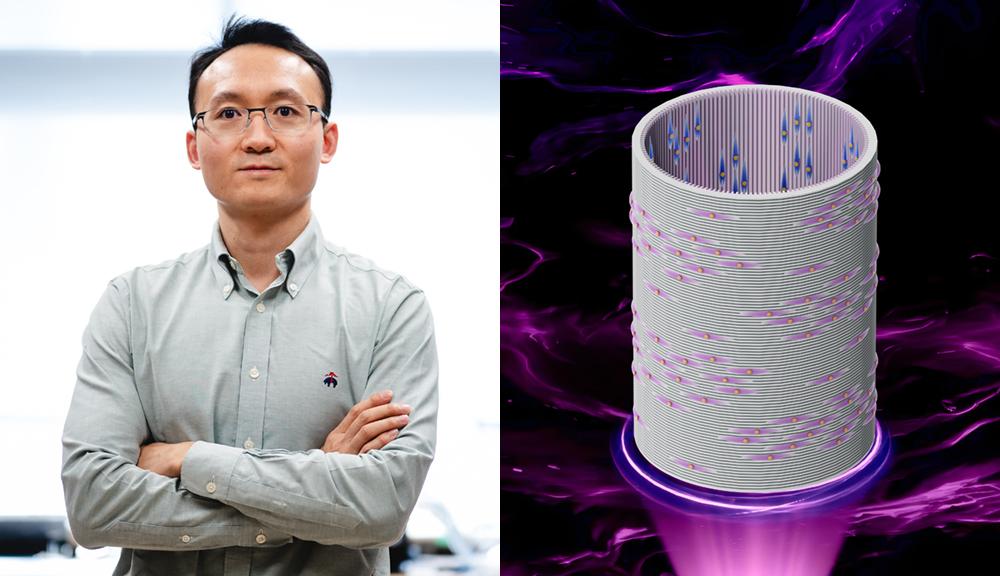 Yonghui Ding, left, and an illustration of a 3D printed blood vessel scaffold. Image via WPI.
Separately, a 3D printed spinal cord implant developed at UC San Diego, reported in 2019, used 200-µm channels to direct neural stem cell growth and encourage axon reconnection in rats. Both assumed a directional relationship between 3D printed geometry and cell behavior.
Neither established which surface features activate which genes or metabolic pathways; without that mechanistic link, geometry cannot be reliably engineered for clinical outcomes. That is the specific gap Owen’s grant funds.
3D Printing Industry is inviting speakers for its 2026 Additive Manufacturing Applications (AMA) series, covering Energy, Healthcare, Automotive and Mobility, Aerospace, Space and Defense, and Software. Each online event focuses on real production deployments, qualification, and supply chain integration. Practitioners interested in contributing can complete the call for speakers form here.
To stay up to date with the latest 3D printing news, don’t forget to subscribe to the 3D Printing Industry newsletter or follow us on LinkedIn.
Explore the full Future of 3D Printing and Executive Survey series from 3D Printing Industry, featuring perspectives from CEOs, engineers, and industry leaders on the industrialization of additive manufacturing, 3D printing industry trends 2026, qualification, supply chains, and additive manufacturing industry analysis.
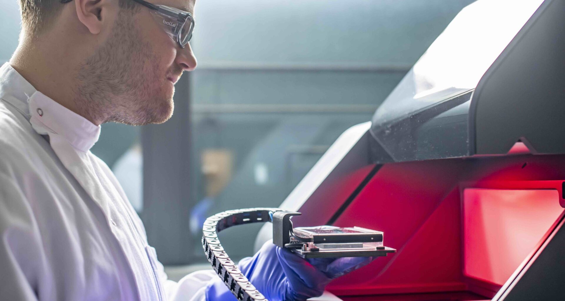
Featured image shows Dr. Robert Owen working with laboratory equipment. Photo via University of Nottingham.