

A new multicenter study suggests that retinal and choroidal imaging may help flag silent brain injury and cognitive risk in people with large artery stenosis (LAS), even when no overt stroke has occurred. Investigators found that OCT and OCT angiography biomarkers were linked to cerebral atrophy and lower cognitive scores, and that adding these eye measurements to clinical models improved prediction of both brain volume loss and cognitive impairment.
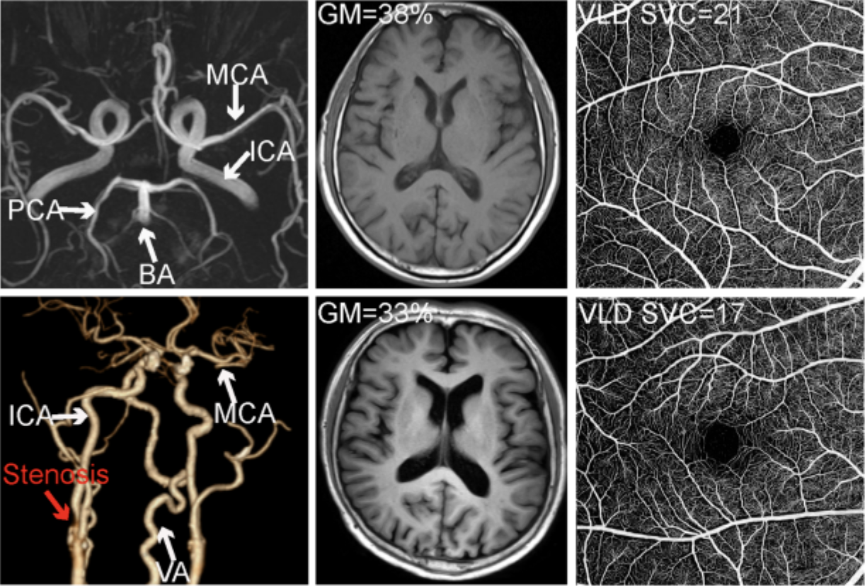
Researchers posit that retinal and choroidal microvascular measures “offer substantial incremental value for individual risk stratification in vascular contributions to cognitive impairment and dementia.” These photos from the study show representative multimodal imaging of a control participant (top) and a nondiabetic patient with noninfarcted large artery stenosis (bottom). CT angiography demonstrates severe carotid artery stenosis in the patient with LAS (red arrow). Photo: Cao L, et al. Invest Ophthalmol Vis Sci. August 3, 2026. Click image to enlarge.
The study, published in Investigative Ophthalmology & Visual Science, examined whether retinal and choroidal microvascular and neurostructural changes reflect brain atrophy and cognitive decline in noninfarcted LAS. The team enrolled 1,239 participants from three tertiary hospitals in China, including 1,035 patients with LAS and 204 age- and sex-matched community controls. The LAS cohort contributed 1,941 eyes, while controls contributed 392 eyes.
Compared with controls, patients with LAS had significantly thinner ganglion cell-inner plexiform layer thickness (63.17μm vs. 66.58μm), lower superficial vascular complex vessel length density (18.64mm-1 vs. 20.69mm-1), lower deep vascular complex vessel length density (26.06mm-1 vs. 27.43mm-1), lower choriocapillaris vessel length density (30.79mm-1 vs. 32.76mm-1) and lower choroidal vascularity index (0.40 vs. 0.43).
Brain volumes were also reduced in patients with large artery stenosis, with lower gray matter/total intracranial volume (35.24% vs. 35.66%) and white matter/total intracranial volume (30.75% vs. 31.37%). Median Montreal Cognitive Assessment scores were lower in patients with LAS than controls (22 vs. 25 out of 30). Retinal and choroidal measures were positively associated with gray matter and white matter volumes, as well as with Montreal Cognitive Assessment scores. These associations were strongest for subcortical regions, especially the thalamus, hippocampus and basal ganglia, and were generally bilateral.
Importantly, the associations between ocular microvascular metrics—particularly choriocapillaris and superficial vascular complex density—and brain/cognitive outcomes were stronger in LAS than in controls, which the authors noted in their paper “suggests that ocular microvascular biomarkers may be more closely linked to neurodegenerative burden in the setting of vascular stress.” However, the team was unable to determine the underlying mechanisms from the study’s cross-sectional data.
Together, the authors wrote that this data “supports retinal and choroidal OCT/OCT-A biomarkers as complementary indicators of vascular-related neurodegeneration relevant to VCID (vascular contributions to cognitive impairment and dementia).” They concluded that these findings “advance the understanding of retina-brain relationships in vascular disease and highlight the potential of retinal and choroidal biomarkers to inform early detection, risk stratification and precision approaches in dementia research.” However, they clarified that future interventional studies are needed “to determine whether improving vascular function modifies ocular biomarkers and downstream brain outcomes.”
Click here for the journal source.
This article was developed by the editorial staff in conjunction with experts in the field. In the process, AI may have been among the editorial tools used to meet the goals of human editors, who approved all content.