
The pace of advancement in myopia management over the past decade has been remarkable. It has been supported by an expanding body of clinical evidence, multiple intervention options and rapidly advancing technology. For busy clinicians, however, this progress brings its own challenges. New studies appear almost weekly, emerging technologies enter the marketplace at an unprecedented rate and commercial messaging can make it difficult to distinguish robust evidence from early promise.
The question that many practitioners face is how to interpret this growing evidence base and translate it into confident clinical decisions. Which interventions have the strongest evidence? How should treatment success be monitored? And how should clinicians balance efficacy with safety when long-term data are still emerging for some approaches?
The International Myopia Institute (IMI) was established to help answer these questions. Bringing together leading experts from around the world across optometry, ophthalmology, vision science and public health, the IMI undertakes comprehensive reviews of the scientific literature and develops consensus reports designed to support evidence-based clinical practice. The emphasis is not simply on summarizing studies but also on critically evaluating the quality, consistency and clinical relevance of the available evidence.
The latest series of IMI white papers highlight a fundamental shift in childhood myopia management from passive correction of refractive error towards proactive identification of risk, prevention where possible, early intervention and personalized treatment strategies aimed at reducing both progression and the lifelong risk of vision-threatening complications. This article summarizes the key clinical messages from the 2025 IMI white papers and provides practical take-home points clinicians can apply in everyday practice.
Getting the Language Right
As myopia management has evolved, so too has the language used to describe it. Terms such as myopia correction, myopia control and myopia management are often used interchangeably in clinical practice, scientific publications and marketing materials. However, the latest IMI definitions editorial argues that these terms describe distinct aspects of care and that greater precision in their use is essential for clear communication among clinicians, researchers, industry, regulators and patients.1
Myopia correction refers to interventions that simply compensate for the refractive error to provide clear distance vision, without any intended effect on slowing myopia progression or reducing axial elongation. Traditional single-vision spectacles and standard contact lenses fall into this category when their sole purpose is to optimize visual acuity.
In contrast, myopia control describes interventions that have been shown to alter the natural course of the condition by slowing refractive progression and excessive eye growth. Many of these treatments also provide refractive correction, but their defining characteristic is evidence demonstrating an effect on myopia progression. The editorial emphasizes that the term should be used thoughtfully and, particularly in marketing contexts, reserved for interventions supported by robust clinical evidence and, where relevant, appropriate regulatory approval.
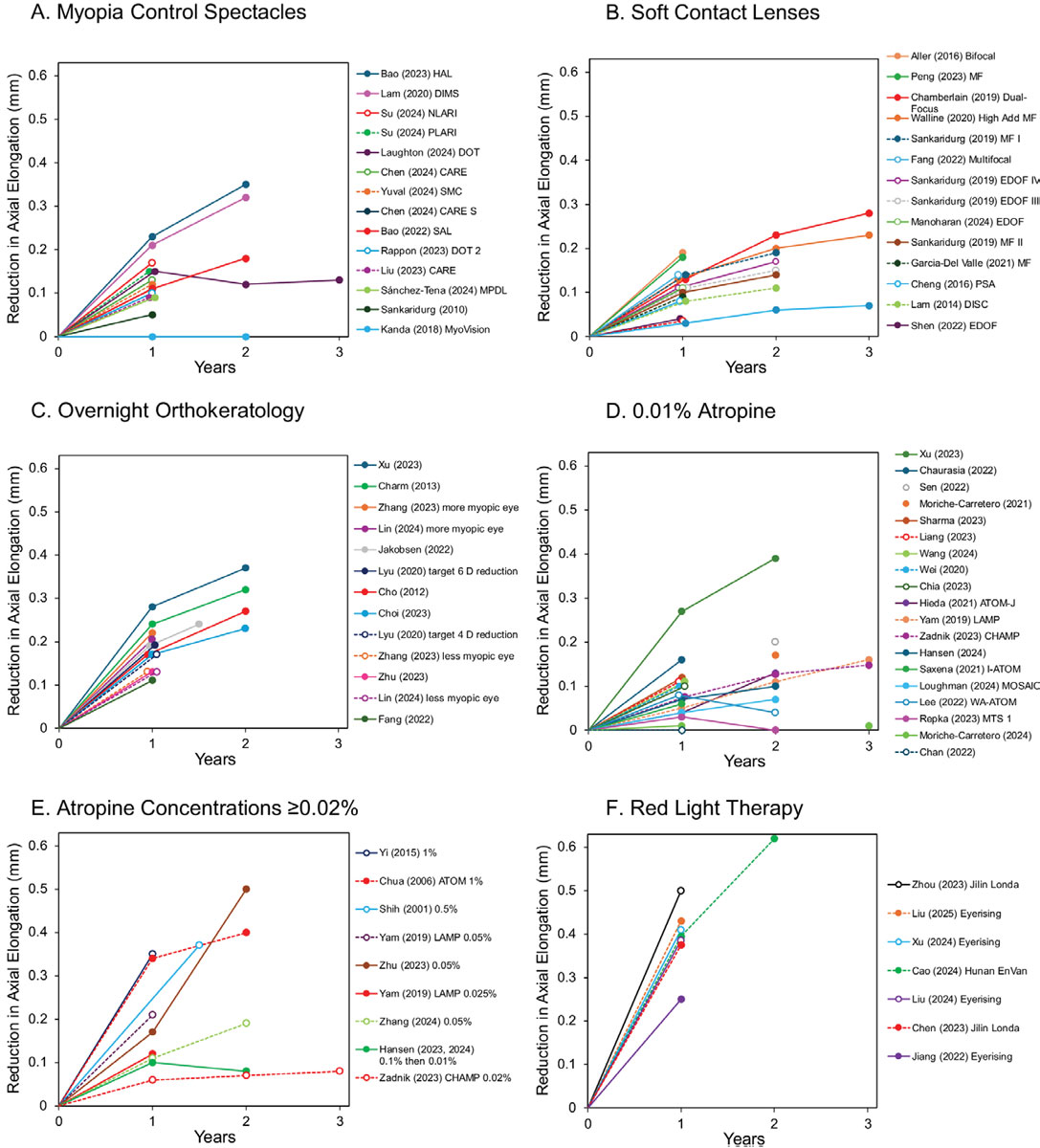
Reduction of axial elongation relative to untreated control children as a function of treatment duration for different myopia control interventions.2 For each panel, the legend is arranged by approximate treatment efficacy, not date: (A) Myopia control spectacles; (B) SoV contact lenses; (C) Overnight orthokeratology; (D) 0.01% atropine; (E) Atropine concentrations ≥0.02%; (F) Red light therapy. Click image to enlarge.
The broadest concept is myopia management. This extends well beyond the choice of treatment and encompasses the lifelong prevention, detection, monitoring and management of myopia and its consequences. It includes identifying children at increased risk of becoming myopic, advising on environmental and lifestyle factors, providing appropriate refractive correction, implementing evidence-based myopia control strategies, monitoring refractive error and axial length (AL) over time and recognizing and managing sight-threatening complications associated with high myopia later in life.
For many decades, clinical care focused largely on correcting blurred vision once myopia had developed. Clinicians are now encouraged to identify children at risk before significant progression occurs, intervene early when appropriate, monitor treatment response objectively and consider the long-term implications of excessive ocular growth.
The terminology we use shapes both professional conversations and patient expectations. Describing care as “myopia management” rather than simply “myopia control” helps parents understand that treatment is not a one-off intervention but a part of an ongoing process that evolves as a child grows and their visual needs change. As the evidence base continues to expand, a shared language provides the foundation for consistent messaging, informed decision-making and high-quality patient care.
The Evidence Base for Interventions Comes of Age
Perhaps the most striking message from the latest IMI interventions review is just how far the field has progressed.2 The debate is no longer whether myopia control works. We now have multiple interventions supported by randomized clinical trials demonstrating meaningful reductions in myopia progression and axial elongation.
In fact, more than 70% of the studies included in this review were published since 2020. For clinicians, this means moving away from a “one-size-fits-all” approach and towards selecting treatments based on the individual child sitting in the consulting room, i.e., their age, rate of progression, lifestyle, risk profile and family preferences.
Importantly, the review only included randomized clinical trials with at least 12 months of follow-up that reported AL outcomes. While refractive error remains important, axial elongation more directly reflects excessive ocular growth and is closely linked to the future risk of retinal detachment, myopic maculopathy, glaucoma and other vision-threatening complications associated with high myopia.
Multiple effective options. One of the most reassuring findings for clinicians is that there is no longer a shortage of effective interventions.
Orthokeratology (ortho-K) remains one of the most consistently effective treatments. Across clinical trials, reductions in axial elongation have approached a median of 0.30mm over two years, with numerous studies demonstrating sustained benefit. For motivated families willing to undertake overnight lens wear, ortho-K continues to be an important option.
Once viewed simply as a means of correcting refractive error, spectacle lenses are now among the widely accepted noninvasive interventions available. Some modern designs have demonstrated reductions in axial elongation of up to 0.35mm over two years, providing an attractive option for younger children and families reluctant to pursue contact lens wear. Myopia control soft lenses have been shown to reduce axial elongation by 0.32mm over a three-year period, making them very comparable with other optical options.
Atropine. This option remains an important component of contemporary myopia management; however, the review reinforces that concentration matters.
Lower concentrations (0.01%) appear to provide modest reductions in axial elongation, while higher concentrations (0.5% or 1%) generally offer greater efficacy. Some studies of higher-dose atropine have reported reductions in axial elongation approaching 0.50mm over two years. Nevertheless, increased efficacy may come at the expense of greater side effects, reduced tolerability and a higher likelihood of rebound once treatment is discontinued. The most appropriate concentration for myopia control is the one that balances efficacy with tolerability. Currently, 0.05% is considered to be the most effective among the lower concentrations prescribed.
Combination therapy. The review found growing evidence that adding low-dose atropine to ortho-K may provide additional reductions in axial elongation compared with ortho-K alone. Although further studies are needed to establish which combinations work best and for whom, clinicians may increasingly consider additive approaches in children who are inherently at high risk and those who continue to progress despite monotherapy.
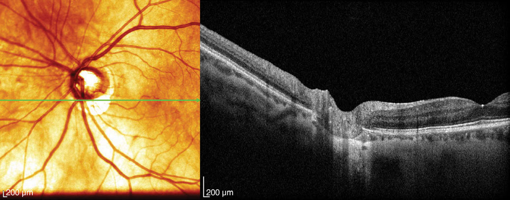
Enhanced depth imaging and swept-source OCT improve visualization of deeper structures, such as the choroid, to reveal thickness and vascular patterns.3 Click image to enlarge.
Promise and precaution. Repeated low-level red-light therapy (RLRL) has generated considerable interest because of the magnitude of its reported treatment effects. Clinical trials have demonstrated median reductions in axial elongation of approximately 0.40mm after one year, exceeding those observed with many established interventions.
Yet the review also urges caution. Reports of reduced foveal cone density and rare cases of vision loss emphasize that efficacy cannot be viewed in isolation from safety. Longer-term studies and ongoing surveillance will be critical before we can fully place this technology within routine clinical practice.
From slowing myopia to preventing it. One of the most exciting developments is the growing focus on preventing or delaying the onset of myopia in children at risk of developing the condition.
Increasing outdoor time remains one of the safest, simplest and most accessible strategies available. The evidence supporting its role in reducing incident myopia continues to strengthen, and encouraging regular outdoor activity should be included in every discussion with families of children at increased risk.
The review also identified emerging evidence that prophylactic treatment may have a future role. A two-year clinical trial demonstrated that 0.05% atropine reduced the likelihood of developing myopia by approximately 50% in children considered at risk. Preliminary evidence also suggests certain optical interventions may eventually contribute to delaying onset, although further research is needed.
What happens when treatment stops? The question of rebound remains highly relevant in clinical practice and is one frequently raised by parents. Encouragingly, current evidence suggests little or no clinically meaningful rebound following cessation of myopia control spectacle lenses or myopia control soft contact lenses. In contrast, higher-dose atropine and repeated low-level red-light therapy appear more susceptible to rebound effects.
Set realistic expectations with families. Treatment plans should include discussions about likely duration of therapy, the possibility of gradual tapering for some interventions and the need for ongoing monitoring after treatment cessation.
Ultimately, the review supports a proactive and individualized approach to care. Clinicians should identify children at risk early, encourage healthy visual behaviors, such as increased outdoor time, monitor refractive error and AL wherever possible and select interventions based not only on efficacy alone. The “best” intervention is not necessarily the one producing the largest treatment effect, but the one that achieves an acceptable balance between effectiveness, safety, tolerability and the needs of the individual child and family.
Measuring What Matters
As myopia management becomes increasingly proactive, the tools we use to assess risk, guide treatment decisions and monitor outcomes have become just as important as the treatments themselves. The latest IMI instrumentation white paper highlights that effective myopia management depends on accurate measurements.3
The starting point remains an accurate assessment of refractive error. Cycloplegic subjective refraction continues to be regarded as the gold standard in children and young adults. However, obtaining a reliable subjective refraction is not always straightforward. When this is not feasible, cycloplegic retinoscopy and cycloplegic autorefraction provide accurate alternatives.
The report reinforces a message that may challenge some established practice patterns: noncycloplegic measurements frequently overestimate myopia because of accommodative influences. Consequently, cycloplegia should be considered an essential component of the examination of children and young adults, extending up to approximately 20 years of age. When cycloplegia cannot be performed, open-field autorefractors appear to provide the closest approximation to cycloplegic findings. The recommended protocols remain familiar, typically one drop of 1% cyclopentolate or two drops of 1% tropicamide administered five minutes apart.
While accurate refraction is fundamental, the most significant shift in recent years has been the growing importance of AL measurement. The interventions review highlighted axial elongation as the preferred outcome measure in myopia control trials; this instrumentation paper reinforces its increasing role in everyday clinical practice.
Modern biometers using partial coherence interferometry, optical low-coherence reflectometry and OCT-based technology allow clinicians to measure ocular growth with remarkable precision and repeatability. Increasingly, AL is becoming the “vital sign” of myopia management.
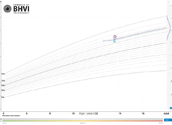
However, the 2025 report also cautions against interpreting AL change in isolation. Some degree of ocular growth is a normal part of child development. The availability of age- and ethnicity-specific growth charts now allows clinicians to compare an individual child’s eye growth against expected population norms. This provides valuable context when evaluating treatment response and helps distinguish normal developmental change from excessive elongation requiring intervention or treatment modification.
Other technologies also play important supporting roles. Corneal topography remains indispensable when fitting and monitoring ortho-K, providing detailed assessment of anterior corneal shape. Corneal tomography extends this evaluation by assessing posterior corneal curvature and corneal thickness, improving screening for potential contraindications. Some instruments also provide information on tear film quality, an increasingly relevant consideration given growing awareness of dry eye symptoms in young patients.
One of the more interesting observations from the report is the renewed attention given to pupil size, which is often overlooked during exams in busy clinical practice. Automated pupillometry provides more reliable measurements than manual assessment and may influence treatment selection and patient counseling. Because optical interventions such as ortho-K and dual-focus contact lenses depend on the interaction between treatment zones and the pupil, pupil size can affect both treatment efficacy and visual quality.
The instrumentation review also reminds clinicians not to lose sight of the posterior segment. Fundus photography and OCT help identify and monitor retinal complications associated with myopia, particularly as patients age and ALs increase. Although choroidal thickness measures continue to generate research interest, their role in clinical decision-making remains uncertain.
Looking to the future, artificial intelligence and predictive analytics may further transform clinical practice. By integrating information such as age, refractive error, AL, family history, environmental exposures and previous treatment response, predictive models may help identify children at greatest risk of progression, estimate future myopia trajectories and guide personalized treatment decisions. Rather than replacing clinical judgment, these technologies could potentially enhance it.
Ultimately, modern instrumentation is no longer simply about documenting baseline findings. The ability to identify risk early, monitor progression accurately and assess treatment response objectively has become fundamental to contemporary myopia management. As our treatment options continue to expand, measuring what matters will be increasingly important in ensuring that the right child receives the right intervention at the right time.
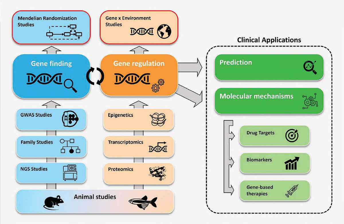
An overview of the current and ongoing myopia-related human genetic research.4 Click image to enlarge.
Moving Towards Personalized Care
While environmental factors such as education, near work and time outdoors have long been recognized as important influences on myopia development, the latest IMI genetics report highlights how genetics contributes to an individual’s risk.4 More importantly, it demonstrates that the future of myopia management is likely to lie in understanding how genetics and the environment interact.
Since the previous IMI genetics report in 2019, research in this area has expanded dramatically. Thousands of genetic variants have now been linked to refractive error and myopia, providing new insights into the biological mechanisms that regulate eye growth and helping explain why some individuals develop high myopia.
One of the clearest messages from the report is that genetics is not destiny. Although myopia has an inherited component, genetic susceptibility interacts closely with environmental exposures. Children who are genetically predisposed to myopia may be particularly vulnerable to factors such as intensive education, prolonged near work and insufficient outdoor time. Increasingly sophisticated analytical methods are strengthening the evidence that these environmental influences can amplify inherited risk.
This growing understanding is driving interest towards more personalized approaches to myopia management. Polygenic risk scores, which combine the effects of multiple genetic variants into a single estimate of risk, show promise as tools for identifying children who may be more susceptible to developing myopia or progressing rapidly. However, the report cautions that these tools are not yet ready for routine clinical use. At present, traditional clinical measures remain more practical and predictive. In particular, cycloplegic refractive assessment continues to be one of the strongest predictors of future myopia risk available to clinicians, with those children with a lower-than-expected hyperopic reserve being more at risk of developing myopia.
Genetic research is also providing valuable insights into the biological pathways involved in myopia development. Large international studies have identified genes involved in retinal signaling, neurodevelopment, regulation of eye size and extracellular matrix remodeling, all of which appear to play a role in ocular growth. Understanding these pathways may ultimately lead to new therapeutic targets and more individualized strategies.
Perhaps one of the most clinically relevant findings relates to children with high or unusually early-onset myopia. There is increasing evidence that some cases may be associated with rare genetic variants linked to inherited retinal disease or connective tissue disorders such as Marfan and Stickler syndromes. Genetic testing has demonstrated a meaningful diagnostic yield, with studies reporting that an underlying genetic condition can be identified in approximately 12% to 23% of cases. In some children, high myopia may be the first or only obvious clinical sign of a broader systemic disorder.
The report also notes growing evidence that myopia shares genetic pathways with other ocular diseases, including retinal detachment, vitreoretinal disorders and primary open-angle glaucoma. Lifelong surveillance in patients with high myopia is important, as management extends well beyond childhood refractive correction.
Family history remains an important component of risk assessment. Children with strong genetic predisposition may benefit from earlier monitoring and intervention, and unusually high or early-onset myopia should prompt consideration of underlying syndromic or inherited disease. Looking ahead, advances in genetics are likely to support increasingly personalized approaches to prevention and treatment. Although genetic testing and risk prediction tools are not yet part of routine clinical practice, the foundations for precision myopia care are rapidly being established.
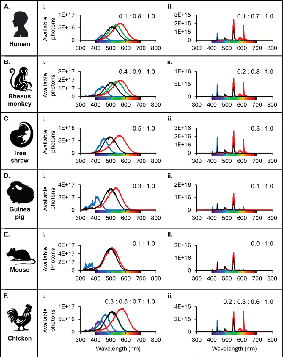
Differences in total photons presented to the retina across species for two different light sources.5 The total photons presented to each classical photoreceptor type were calculated between 300nm and 800nm. Click image to enlarge.
Revisiting Environmental Influences
If the genetics report highlights who may be at greater risk of developing myopia, the latest IMI white paper on light explores what may be one of the most important environmental influences that can modify that risk.5 Although outdoor activity has been recommended as a cornerstone of myopia prevention for many years, researchers are still working to understand precisely why spending time outdoors appears to protect against myopia development.
The 2025 IMI light report examines the growing body of evidence investigating how different characteristics of light (including intensity, spectral composition and timing) may influence eye growth and refractive development. Together, these findings provide important biological support for many of the clinical recommendations already being made in practice. Much of our current understanding comes from animal models, where light has consistently been shown to influence refractive development. One of the most compelling findings is the role of retinal dopamine, a neurotransmitter that appears to act as a natural regulator of eye growth. Exposure to bright light stimulates dopamine release within the retina, and increased dopamine activity has been associated with reduced axial elongation. Researchers have proposed that dopamine may function as a biological “brake” that helps protect against excessive eye growth and the development of myopia.
Animal studies have also demonstrated that changes in spectral composition, exposure to specific wavelengths and disruption of normal light-dark cycles have all been shown to alter eye growth. While these findings provide important insights into the biology of myopia, the report cautions that direct translation to humans remains challenging because of substantial differences between species in retinal structure, visual processing and ocular development.
In humans, the relationship between light exposure and myopia is more complex. Although a growing body of epidemiological evidence suggests that children who spend more time outdoors are less likely to become myopic, it remains difficult to determine exactly which aspects of the outdoor environment are responsible for this protective effect. Brighter light exposure is one likely contributor, but outdoor environments also differ from indoor environments in terms of viewing distances, visual complexity, spatial frequency content and spectral composition.
Nevertheless, evidence from wearable light sensors and population studies continues to support the notion that brighter daily environments are associated with a lower risk of developing myopia. Importantly, the protective effect of outdoor activity has now been demonstrated across multiple populations and remains one of the most consistent findings in myopia research.
Emerging areas of interest include sleep patterns, circadian rhythms and exposure to artificial light at night. While some studies suggest potential associations between sleep disruption, altered sleep timing and myopia development, current evidence remains inconsistent. At present, there is insufficient evidence to conclude that sleep patterns directly influence refractive development, although ongoing research continues to investigate possible links between circadian regulation and ocular growth.
For clinicians, perhaps the most important message is that the practical advice remains remarkably straightforward despite increasing scientific complexity. Evidence continues to support encouraging children to spend at least two hours outdoors each day as part of a comprehensive myopia prevention strategy. Parents can also be reassured that this benefit appears to persist under typical outdoor conditions, including cloudy weather, and is not negated by using sunglasses or other standard forms of ultraviolet protection.
The report also considers emerging light-based interventions such as repeated low-level red-light therapy. While clinical studies have demonstrated impressive reductions in axial elongation, the report advocates a cautious approach until further long-term evidence becomes available.
Myopia development is shaped by a complex interaction between genetics, environment and behavior. While researchers continue to unravel the underlying biological mechanisms, the clinical message remains clear. Encouraging regular outdoor activity remains one of the simplest, safest and most accessible interventions available for reducing the risk of myopia onset, while advances in our understanding of light-mediated pathways may help inform future preventative and therapeutic strategies.
The IMI Digest 2025
One of the challenges facing clinicians today is not a lack of information, but the sheer volume of it. Thousands of myopia-related papers are published each year, making it increasingly difficult for busy practitioners to identify which findings are clinically meaningful, which are supported by robust evidence and which remain preliminary. To address this challenge, the IMI publishes a biennial Digest that provides concise updates to previously published white papers in areas where significant advances have occurred.6
The IMI 2025 Digest highlights six areas in which the evidence base has evolved substantially: definitions and classification of myopia, clinical management, risk factors, accommodation and binocular vision, experimental models of myopia and myopia onset and progression in young adults. Rather than revisiting entire topics, the Digest focuses on developments that are most relevant to clinical practice.
A major theme emerging from the 2025 Digest is the growing emphasis on prevention and earlier intervention. The concept of pre-myopia continues to evolve, with increasing attention being paid to hyperopic reserve as an important predictor of future myopia development. This refers to the age-appropriate level of hyperopia that offers a protective buffer against the development of myopia, with higher levels required at younger ages to reduce the risk. Population differences may influence the amount of hyperopic reserve required to reduce risk, with Asian populations potentially requiring a greater hyperopic reserve to avert the risk of myopia onset.
The Digest also reinforces that delaying myopia onset may be one of the most powerful strategies available to reduce lifetime myopia burden—a key message that has become increasingly prominent across the myopia literature. Delaying onset by even one year may have a greater impact on final refractive error than several years of subsequent myopia control treatment.
In the area of risk factors, the Digest highlights important methodological advances that are helping researchers move beyond simple associations and better understand causality. New population research approaches continue to strengthen evidence supporting the roles of education and time outdoors in myopia development. At the same time, the report notes that evidence linking factors such as screen use and sleep remains inconsistent, emphasizing the importance of interpreting emerging research cautiously.
Updates on accommodation and binocular vision provide reassurance for clinicians, confirming that modern myopia control interventions have minimal long-term impact on general accommodative and binocular function and changes in these measures are not consistently associated with myopia progression. This supports the continued use of evidence-based myopia control treatments without concern that they may adversely affect binocular visual function.
The Digest also summarizes important advances from experimental models of myopia. Research is increasingly uncovering the biological mechanisms that regulate eye growth, including the role of light exposure, retinal signaling pathways, novel opsins and molecular processes within the choroid and sclera. Although much of this work remains at the laboratory stage, these discoveries are helping explain clinical observations and may ultimately lead to new therapeutic approaches.
Finally, the Digest provides further evidence that myopia progression does not necessarily stop at the end of adolescence. New longitudinal studies confirm that a proportion of young adults, particularly those with higher levels of myopia, longer AL or intensive educational and occupational demands, continue to experience clinically meaningful progression. Myopia management should be viewed as a lifelong process rather than a pediatric intervention alone.
Collectively, the IMI 2025 Digest reflects a field that continues to evolve rapidly. Myopia management is increasingly shifting toward earlier identification, individualized care and lifelong risk reduction. For clinicians, the Digest provides a practical and evidence-based overview of the developments most likely to influence patient care while helping bridge the gap between emerging research and everyday clinical practice.
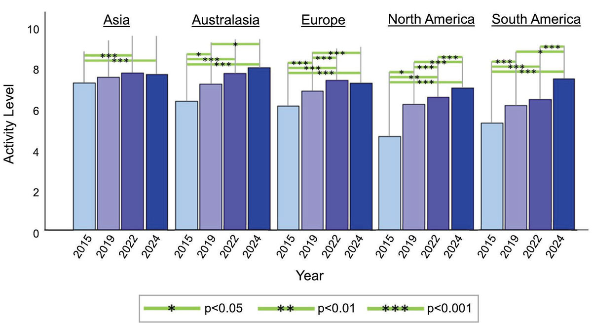
Changes in practitioner’s perceived level of clinical activity in myopia management by continent between 2015 and 2024. Green horizontal lines between years indicate statistically significant change and no line indicates no significant change.7 Click image to enlarge.
How Clinical Practice Is Evolving Worldwide
To better understand how practitioners are responding to emerging evidence, the IMI has conducted a series of global surveys since 2015, tracking attitudes, prescribing behaviors, access to diagnostic technology and perceived barriers to care. The latest survey, completed in 2024, provides a unique nine-year perspective on how myopia management is evolving across different regions of the world.7
Concern about childhood myopia continues to increase globally. Practitioners in Asia and South America reported the highest levels of concern regarding the condition’s growing prevalence in children, while concern was somewhat lower, although still substantial, in Australasia. Compared with earlier surveys, concern has increased significantly across most regions, reflecting growing awareness of the long-term ocular health consequences associated with progressive myopia.
The survey also highlights important changes in clinical management patterns. Although single-vision spectacles remain the most commonly prescribed correction for myopic children globally, their use has steadily declined over the past decade. At the same time, prescribing of evidence-based myopia control interventions, including specialized spectacle lenses, soft contact lenses, ortho-K and low-dose atropine, has increased substantially. These trends suggest that clinicians are increasingly moving beyond simple refractive correction and embracing proactive myopia management strategies.
Access to diagnostic instrumentation continues to vary considerably around the world. While autorefraction and keratometry are widely available, access to cycloplegic refraction and AL measurement remains less consistent. Encouragingly, practitioners are initiating treatment earlier than they were a decade ago. The minimum level of myopia and the amount of annual progression required before commencing treatment have both decreased over time, reflecting increasing acceptance of the benefits of early intervention and risk reduction. This trend aligns closely with the themes emerging throughout the IMI 2025 reports, particularly the growing emphasis on prevention, pre-myopia and proactive management.
Undercorrection, once proposed as a potential strategy for slowing progression, has now been largely abandoned, with more than 80% of practitioners reporting that they never use it. This reflects the profession’s increasing reliance on evidence-based approaches and highlights the value of consensus guidance in helping clinicians navigate an expanding evidence base.
Despite these advances, important barriers remain. Cost continues to be the most reported obstacle to prescribing myopia control interventions, followed by limited treatment availability and lack of practitioner information or training. These findings highlight the ongoing need for education, improved access to care and the development of affordable treatment options if the benefits of myopia management are to be realized on a broader population level.
Perhaps most notably, practitioners report that incorporating myopia management into clinical practice delivers benefits beyond patient outcomes alone. Many clinicians indicated improvements in patient loyalty, professional satisfaction and practice growth. This suggests that evidence-based myopia management is increasingly being viewed not as a niche service, but as a core component of contemporary eye care.
Taken together, the survey findings paint a picture of a profession in transition. Awareness of myopia as a significant public health issue continues to grow, clinicians are adopting evidence-based interventions earlier and more frequently and management is increasingly centered on prevention and long-term risk reduction. Although challenges relating to access, affordability and education remain, the global direction of travel is clear: myopia management is becoming an integral part of routine clinical practice worldwide.
From Correcting Blur to Managing Lifelong Risk
The 2025 IMI white papers collectively illustrate just how far myopia management has evolved. What was once largely limited to correcting refractive error has become a sophisticated, evidence-based field focused on prevention, early intervention, objective monitoring and long-term risk reduction. Around the world, clinicians are intervening earlier, adopting evidence-based treatments more frequently and increasingly viewing myopia management as a core component of contemporary eye care rather than a niche area of practice.
Perhaps the most important message from the 2025 IMI series is that myopia management should be viewed as a lifelong process. The goal is not simply to reduce refractive progression during childhood but also to minimize the cumulative burden of axial elongation and ultimately reduce the risk of vision-threatening complications later in life.
As the volume of myopia research continues to expand, the challenge for clinicians will be distinguishing meaningful advances from emerging hypotheses and translating evidence into practical patient care. By bringing together international experts to critically evaluate the highest-quality evidence and develop consensus recommendations, the IMI continues to provide a trusted framework to support evidence-based decision-making. For clinicians, this means greater confidence that the recommendations implemented in practice are grounded not only in the latest research but also in a rigorous assessment of what that research means for patients.
Dr. Jones is a clinical professor at the School of Optometry and Vision Science and a Lead clinical scientist at the Centre for Ocular Research & Education (CORE), at the University of Waterloo. She is a fellow of the British College of Optometrists, the British Contact Lens Association and the American Academy of Optometry. She has no financial disclosures.
Dr. Tahhan is executive director of the International Myopia Institute and an optometrist and researcher with expertise in myopia, global eye health and evidence-based clinical practice. She is a consultant at the Brien Holden Vision Institute and senior conjoint lecturer at the School of Optometry and Vision Science at the University of New South Wales. She has no financial disclosures.
Dr. Wolffsohn is a professor of optometry based at Aston University in Birmingham in the United Kingdom. He is the chief scientific officer of the International Myopia Institute and a consultant to Alcon, Bausch + Lomb, Coopervision, DopaVision, Essilor, Santen, SightGlass Vision and Topcon.
Dr. Voogelaar completed medical school before joining the department of ophthalmology at Erasmus University Medical Center and the Rotterdam Eye Hospital as a PhD candidate. He has no financial disclosures.
Dr. Harb is a clinical professor at the Herbert Wertheim School of Optometry at the University of California at Berkeley and in the department of ophthalmology at University of California at San Francisco. Her financial disclosures include CooperVision and Meta Reality Labs (research funding).
Dr. Ashby is associate professor of neuroscience and director of graduate research in the faculty of science and technology at the University of Canberra, Australia. He is also an adjunct associate professor at The Australian National University and the University of New South Wales, Australia. He has no financial disclosures.