
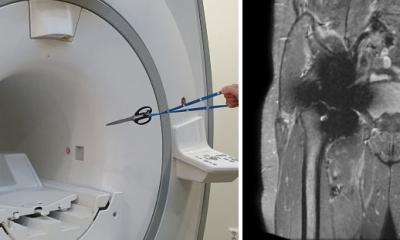
A previous MRI does not ensure patient safety in any way for future MRI examinations
Christos Tsiotsios
The less is known about an implant, the more cautious radiographers need to be, Tsiotsios advised, describing his approach as an “identification pyramid”: starting with the implant card and manufacturer’s website as the gold standard, then working down through electronic health records, surgical notes, specialist databases such as MRIsafety.com or MagResource.com, prior imaging, and finally plain radiography or CT. Crucially, he warned, ‘a previous MRI does not ensure patient safety in any way for future MRI examinations.’ The expert singled out intracranial aneurysm clips as a particular category of concern: scanning must not proceed until the specific manufacturer, model, and type have been confirmed, as the consequences of error can be fatal.10 ‘For unknown passive implants, the policy should be a collaborative effort between the supervising physician, the radiographer, and the medical physicist,’ Tsiotsios advised.
Looking ahead, the expert discussed the potential of artificial intelligence to assist with pre-examination implant identification by analysing electronic health records and prior imaging, with studies already reporting accuracy above 80%.11 While these AI system show great promise for enhancing workflow efficiency and MRI safety, data privacy and validation challenges must be addressed, and human oversight remains essential, he concluded: ‘The label provides the limits, but the clinical team ensures patient safety.’
Profiles:
Dr Simone Busoni is a senior medical physicist at the UOC Medical Physics department of Careggi University Hospital in Florence, Italy, where he holds a Level III Radiation Protection Expert qualification — the highest certification level in Italy. He serves as coordinator of the EFOMP working group responsible for revising Policy Statement 14 on MRI Safety, the European reference framework for safe MRI practice, and is a member of the ESR Radiology MR Safety and Quality Subcommittee.
Prof. Siegfried Trattnig is Professor of Radiology with a specialist focus on high-field MRI at the Medical University of Vienna, Austria. Since 2000, he has served as Medical Director of the University of Vienna’s high-field MR research scanner, and since its founding in 2003, as Director of the High Field MR Centre of Excellence at the Medical University of Vienna. He has been a member of more than 50 committees across the major international societies in radiology, orthopaedics, and MR imaging, including the ESR Research Committee Board; he has served as Chairman of the European Imaging Biomarker Alliance of the ESR, Chairman of the ISMRM Musculoskeletal Study Group, and Director of the School of MRI of the ESMRMB. He sits on the editorial boards of seven peer-reviewed journals and has authored over 500 indexed publications in PubMed as well as 24 book chapters.
Edwin H.G. Oei, MD, PhD, is Associate Professor of Musculoskeletal Imaging and Section Chief of Musculoskeletal Radiology in the Department of Radiology and Nuclear Medicine at Erasmus MC, University Medical Center, Rotterdam, the Netherlands. As principal investigator of the Advanced Musculoskeletal Imaging Research group at Erasmus MC (ADMIRE), his research focuses on multi-parametric MRI, CT, and ultrasound imaging of diseased joints. His work encompasses the technical development, validation, and implementation of novel MR techniques in large clinical and population-based studies.
Christos Tsiotsios, MSc, is Head of Radiographers at Markides MRI and Diagnostic Imaging in Limassol, Cyprus, and a part-time instructor at the European University of Cyprus in Nicosia. He specialises in MR safety, with a particular focus on the practical management of patients with passive implants in clinical radiography practice.
References:
Nordin LE, Åberg K, Kihlborg J et al.: ESR Essentials: basic physics of MR safety—practice recommendations by the European Society for Magnetic Resonance in Medicine and Biology; European Radiology 2024 European Society of Radiology (ESR): The European MR safety landscape; Insights into Imaging 2024 Pogliacomi F, Schiavi P, Calderazzi F et al.: Is there a relation between clinical scores and serum ion levels after MoM-THA? One year results in 383 implants; Acta Biomedica 2020 Bruschetta A, Palco M, Fenga D et al.: How to Manage Metallosis: A Retrospective Cohort Analysis after Revision Hip Surgery; Journal of Clinical Medicine 2023 Feuerriegel GC, Sutter R: Managing hardware-related metal artifacts in MRI: current and evolving techniques; Skeletal Radiology 2024 Kim Yj, Yoon D, Song SO, Lee S, Choo HJ, Ryu J: Metal Artifacts in Postoperative Spine MRI: Strategies and Limitations; Investigative Magnetic Resonance Imaging 2025 Luitjens J, Ziegeler K, Yoon D et al.: Improved metal suppression using new generation low-field MRI: a biophantom feasibility study; Skeletal Radiology 2024 Aboelmagd S, Malcolm P, Toms A: Magnetic resonance imaging of metal artifact reduction sequences in the assessment of metal-on-metal hip prostheses; Reports in Medical Imaging 2014 Ziegeler K, Yoon D, Hoff M, Theologis AA: Metal Suppression Magnetic Resonance Imaging Techniques in Orthopaedic and Spine Surgery; Journal of the American Academy of Orthopedic Surgeons 2025 Klucznik RP, Carrier DA, Pyka R, Haid RW: Placement of a ferromagnetic intracerebral aneurysm clip in a magnetic field with a fatal outcome; Radiology 1993 Valtchinov VI, Lacson R, Wang AW, Khorasani R: Comparing Artificial Intelligence Approaches to Retrieve Clinical Reports Documenting Implantable Devices Posing MRI Safety Risks; Journal of the American College of Radiology 2020