
 Credit: luismmolina/Getty Images
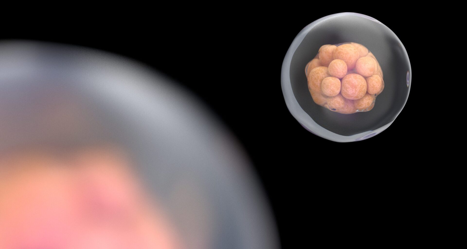
Successful fertility treatments, including in vitro fertilization (IVF), is often reliant on a clear understanding of developmental stages of human development. Clinicians working with assisted conception technologies rely on a variety of tests to assess the viability of embryos prior to implantation.
Researchers at the Loke Centre for Trophoblast Research at the University of Cambridge have been studying development of human pre-implantation embryos in an effort to improve success of embryo growth and development to the stage where they might be transferred.
“Having a baby through assisted conception can be very challenging,” said senior author Kathy Niakan, PhD, director of the Loke Centre for Trophoblast Research. “Most embryos fail to develop or to implant, and even those that are good quality may not be transferred. Much more basic research is needed to inform future clinical practice and improve rates of assisted conception.”
Niakan and her team of researchers at the Loke Centre and Francis Crick Institute developed a technique to observe embryo development in real time at high resolution, while also monitoring for genetic abnormalities that may arise.
Their work was published in Nature Biotechnology in a paper entitled, “Live imaging of late-stage preimplantation human embryos reveals de novo mitotic errors.”
Using an imaging technique called light-sheet microscopy, embryos donated by families with successful pregnancies were observed in 3D without damage to the embryos.
“This is the first time that this very gentle method, with much higher resolution, has been used,” shared first author Ahmed Abdelbaki, PhD, a postdoctoral researcher at the Loke Centre for Trophoblast Reseach. “It meant that we could watch the embryos as they developed over a two-day period, the longest continuous time that this process has been observed.”
The authors described in their paper how they systematically tested multiple nuclear labeling methods and eventually developed a method for electroporation to introduce tagged mRNA into the embryos. They wrote, “To trace individual nuclei, we developed an open-source, semi-automated segmentation method using a customized deep learning model optimized for variability in embryo size, shape and signal.”
“The unique design of the microscope allows for multiple precious embryos to be watched simultaneously, and from both sides. This has allowed the team to catch events that have previously been missed,” said co-author Ben Steventon, PhD, from the University of Cambridge.
Using this approach, the team identified that the majority of cells developing genetic abnormalities, including de novo multipolar divisions, lagging chromosomes, misalignment and chromosome slippage occurred in cells on the exterior of the embryo—cells that typically develop into the placenta.
Approximately 10% of the 13 embryos utilized in this study contained chromosomal abnormalities. While these abnormalities are tolerated in the pre-implantation embryos, more research is needed to identify the long-term fates of these cells.
“We were extremely surprised to discover that abnormal cell divisions can happen from scratch at a very late stage of human development,” said Niakan. “It was only by using a new imaging technique that it was possible to see this happening.”
“It’s a proof of the power of direct observation to uncover unexpected findings in human biology,” Steventon commented.
This discovery is relevant for the future of assisted conception technologies, as the very cells that have been observed to have genetic abnormalities, which are often fated to become placental, are the cells that are typically biopsied for use in preimplantation genetic testing. Their work raises question about the clinical utility of this testing to determine embryo health.
“Our data strongly advocate for further research into the underlying cause and consequence of late-stage aneuploidies. Moreover, our findings suggest reconsideration of embryo transfer timing, because in vitro culture increases the risk of chromosome segregation errors,” the authors concluded.