
Over 180 million years, microbes inside a decaying ichthyosaur, an extinct marine reptile, actively transformed its skeletal remains into a stunning, three-dimensional fossil. This discovery reshapes the way scientists understand the fossilization process, showing that it is not just an external environmental phenomenon, but an intricate interaction between life and death, chemistry and biology. The new insights open doors to rethinking how fossils are preserved and could guide future searches for ancient life, not only on Earth but possibly beyond.
The Discovery: Microbes at the Core of Fossilization
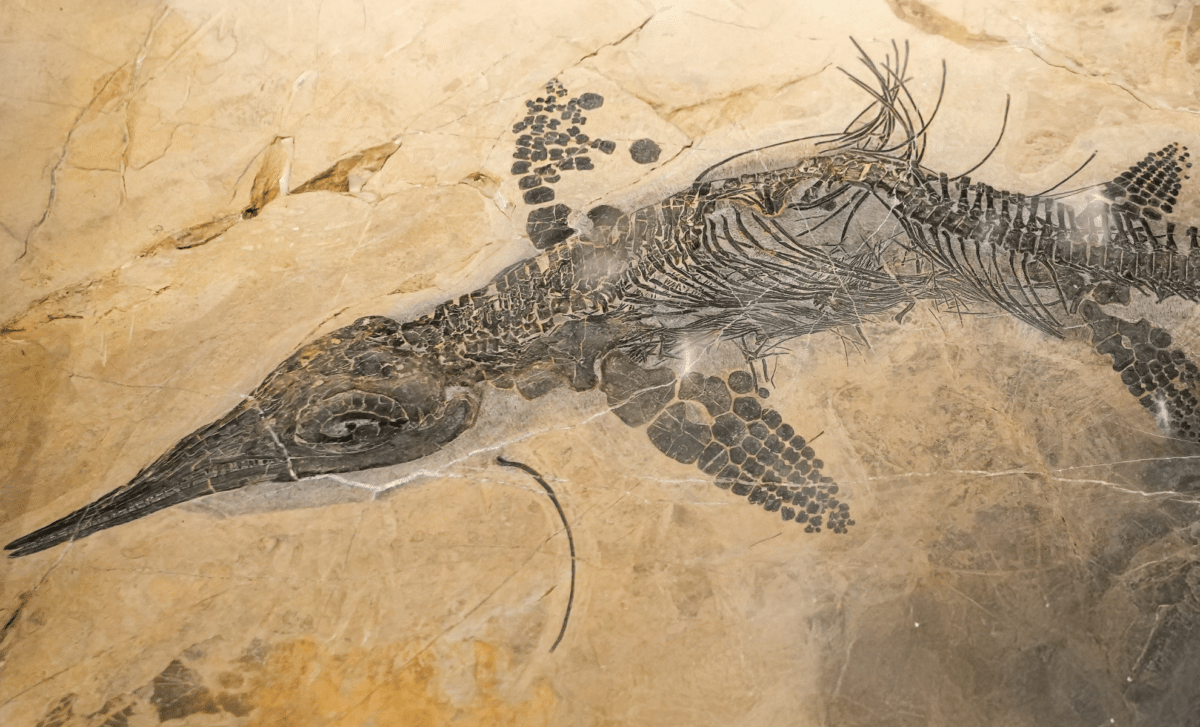
The ichthyosaur fossil in question, discovered in southwestern Germany, has been preserved in exceptional three-dimensional detail. This breakthrough study, led by Andrew Jian, a doctoral researcher at Curtin University, overturns the traditional view that fossilization is solely a process controlled by environmental factors like the surrounding seafloor. Instead, Jian’s team has shown that microbes inside the decaying animal played an essential role in its preservation. The study, published in Nature Communications, suggests that the fossilization process is far more dynamic, driven not only by external conditions but also by microbial activity inside the body.
Jian’s team used advanced techniques to examine the bones of the ichthyosaur, which were encased in a carbonate nodule. The team discovered three distinct chemical zones within the fossil, each of which provided crucial clues about the animal’s decay process and subsequent mineralization. By studying these zones, Jian identified how microbial activity contributed to the mineral structure inside the bones, preserving their three-dimensional shape. The study highlights that microbes released acids during decomposition, which allowed minerals like barite to form before the bones were sealed by calcite and other minerals. This early mineralization protected the bones from external pressure as sediments built up around them over millions of years.
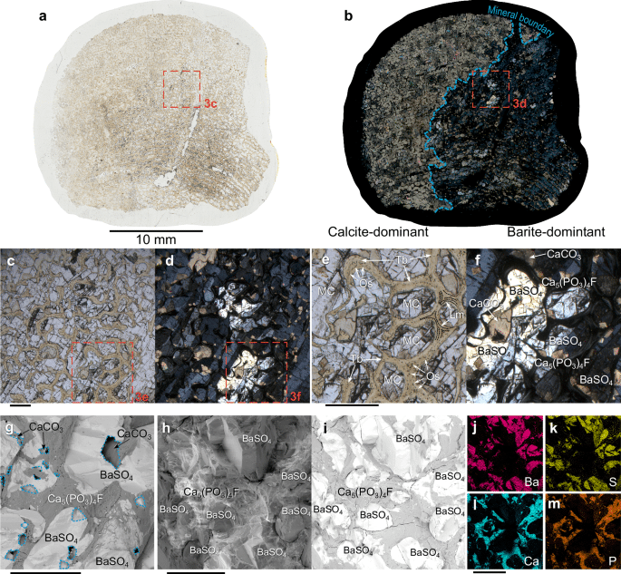 Microscopic mineralogy in ichthyosaur vertebra.
a Thin section map of 25 mm cored vertebra sample Vert-E under plain polarized light (PPL). The thin section was sliced through the transverse plane of the vertebra. b Map a under cross polarized light (XPL). A mineral boundary (blue dashed line) between calcite infilling peripheral cavities and barite filling the interior cavities. Scale bar for a, b 10 mm. c, Micrograph of spongy bone trabeculae and marrow cavities under PPL. d Same view of c under XPL. e Inset of c. Tb = trabeculae, MC = marrow cavities, Lm = lamellae (black dashed lines), Os = osteocytes (dark spots). f Inset of d with identified minerals. Barite = BaSO4, calcite = CaCO3, fluorapatite = Ca5(PO3)4F. g Backscatter electron (BSE) micrograph of Vert-B with identified minerals. Voids marked in blue dotted lines were calcite crystals dissolved during acid treatment. h Secondary electron (SE) micrograph of Vert-D. i, BSE view of h. Energy Dispersive X-ray Spectroscopy (EDS) maps of view (h, i), for j barium, k sulfur, l calcium and m phosphorous. j and k indicate barite in marrow cavities, l, m indicate fluorapatite composing trabeculae. Scale bar for c–m 500 μm. Larger thin-section maps are shown in Supplementary Fig. 6a, b.
The Role of Microbes in Preserving Fossils
As the ichthyosaur decayed, two distinct groups of microbes carried out different functions inside the body. One group broke down organic matter, while another likely pushed sulfur back toward sulfate, aiding in the preservation of the skeleton. The researchers found that these microbial actions happened within small pockets, where chemical reactions could take place in isolation from the oxygen-starved seafloor environment. This allowed the body to remain intact, preserving its shape and structure for millions of years.
Jian explained, “What we found shows the fossilisation story is far more complex.” These microbes, often invisible to the naked eye, were not mere passive participants in the decay process. They actively contributed to the formation of minerals, ensuring that the bones retained their three-dimensional form even as soft tissues broke down. This finding challenges traditional ideas of fossilization and reveals the intricate, interconnected processes that can lead to the exceptional preservation of ancient organisms.
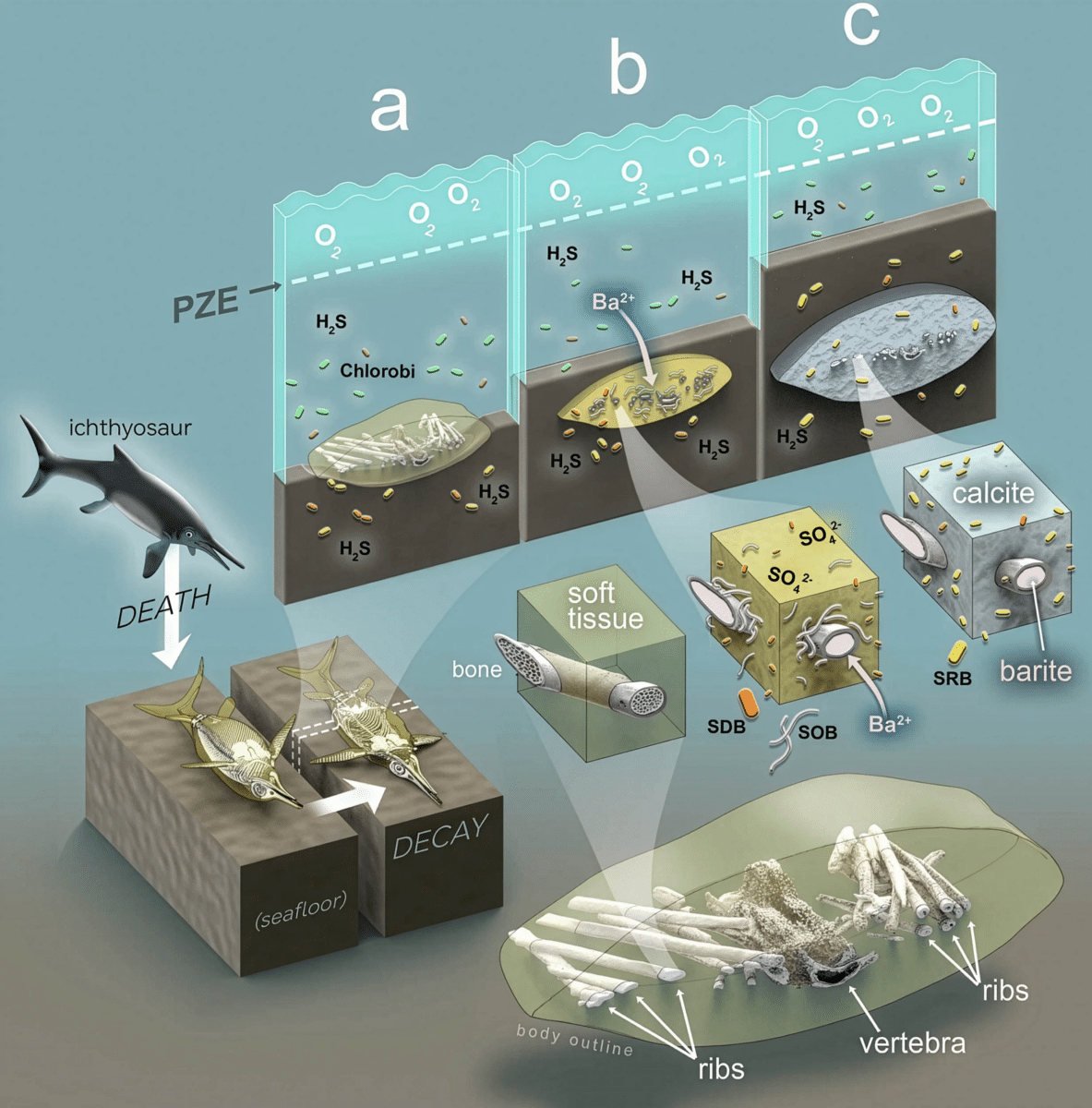 Graphical representation showing how microbes helped create this 3D ichthyosaur fossilization sequence.
Credit: Nature Communications Earth & Environment.
The Environment: A Hostile Yet Preservative Setting
The fossil was found in the Posidonia Shale formation, a geological site known for its lack of oxygen and high concentration of sulfides. This euxinic environment, rich in sulfur and poor in oxygen, would have been a difficult place for larger scavengers to survive. However, despite the hostile conditions, the ichthyosaur’s remains were able to retain their structure due to the unique chemical environment created by the microbes inside its body. The sulfide-rich waters also left chemical fingerprints that helped researchers trace the microbial processes that occurred during the decay and fossilization stages.
The discovery that microbes could preserve an organism’s shape in such an oxygen-deprived environment is revolutionary. It shows that even in the harshest conditions, the decay process can lead to the formation of fossils, albeit through a more complex set of interactions than previously thought. This insight into ancient ecosystems provides an invaluable understanding of the conditions under which rare and exceptional fossils might form.