
For centuries, the inner surface of the human heart concealed one of anatomy’s quiet riddles. Covered in a complex, lace-like network of muscular strands called trabeculae, the heart’s interior had been observed and drawn in extraordinary detail by Leonardo da Vinci, yet their function in adult humans remained, for the longest time, entirely unknown.
Da Vinci, who studied the heart through direct dissection, theorized that the branching, snowflake-like structures served as a kind of warming network for blood, not unlike the systems used today to prevent roads and sidewalks from freezing.
That theory, compelling as it was, went unverified for five centuries. It wasn’t until a team of researchers published their findings in the journal Nature in 2020 that the scientific community finally had answers, and they came from an unlikely combination of disciplines.
A Study of Unprecedented Scale
To crack the mystery, scientists turned to the UK Biobank, analyzing data from 18,096 participants. According to the research published in Nature, the inner surfaces of the human heart are covered by a complex network of muscular strands thought to be a remnant of embryonic development, and until this study, both the function of those trabeculae in adults and their genetic architecture were unknown.
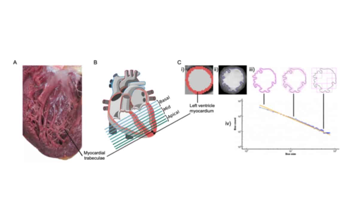 Figure showing exploration of human heart physiology – © Nature
Using biomechanical simulations alongside observational data drawn from human participants, the team was able to demonstrate that trabecular morphology, the specific shape and structure of this branching network, is an important determinant of cardiac performance. In practical terms, that means certain trabecular configurations were linked to an increased risk of cardiovascular disease, a finding that carries real weight for clinical medicine.
Fractal Theory Meets Cardiac Science
The analytical backbone of the study was fractal analysis, a mathematical framework that describes how patterns repeat at different scales, from Google Maps photographing entire countries through progressively smaller aerial tiles, to tree branches splitting into ever-finer limbs, to telecommunications networks blanketing the Earth. In the case of the heart, the trabeculae branch into smaller and smaller threads, and it is precisely the nature of that fractal network where many of the key clues were found.
According to Popular Mechanics, the researchers were able to identify common structural features across different patient imagery, then draw conclusions about what those structures are doing functionally. The use of MRI technology was central to capturing the detail required for this kind of analysis at such a large scale.
Genetics Adds Another Layer
Beyond anatomy and imaging, the study went a step further into the genome. The researchers identified 16 significant loci, specific locations in the genetic code, containing genes associated with haemodynamic phenotypes and the regulation of cytoskeletal arborization. Arborization, worth noting, is simply the technical term for branching, as in the structure of a tree.
By cross-referencing the genetic data of tens of thousands of subjects with their trabecular structures, the team began to identify the genomic mechanisms that govern how trabeculae form and function, findings that may hold implications for understanding how other types of body cells develop and behave as well.
As researcher Hannah Meyer put it in a statement: “Only the combination of genetics, clinical research, and bioengineering led us to discover the unexpected role of myocardial trabeculae in the function of the adult heart.” The researchers themselves acknowledge this is only a first step toward a more complete understanding of these structures.