
New ligase ribozymes isolated from in vitro evolution
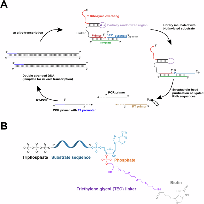
Recently, we used in vitro evolution to ask whether and how ligase ribozymes might switch substrate specificity from RNA oligonucleotides activated with a 5′-phosphorimidazole group (5′-AIP) to those containing the biologically relevant 5′-triphosphate group (5′-PPP)11. In that work, we created a partially randomized RNA library derived from a previously characterized ‘phosphorimidazolide ligase′ (AIP-Ligase)12 by mutagenizing a 40-nt region of this ribozyme at 21% per nucleotide position. This variable region was flanked by constant regions, which provide binding sites for reverse transcription and PCR primers. The 3′ constant region included an 8-nt ‘primer′ sequence connected to the rest of the construct by a hexauridine linker. The terminal nucleotide of this primer sequence (i.e., the last nucleotide of the selection construct) was the intended site of ligation (Fig. 1A). Library sequences with the ability to ligate to a 16-nt RNA substrate containing a 5′-triphosphate group and 3′-biotin tag were purified away from unreactive sequences by streptavidin bead capture and subsequently amplified by RT-PCR and in vitro transcription (Fig. 1). The substrate sequence was connected to a triethylene glycol (TEG) linker by its 3′-phosphate group, and the TEG linker was in turn covalently attached to a biotin group (Fig. 1B). The target reaction for this selection involved a nucleophilic attack of the 3′-hydroxyl (3′-OH) end of the selection construct (the ‘primer′) on the α-phosphate of the 5′-PPP group of the substrate. (Fig. 1). Although we isolated five distinct classes of ligase ribozymes from that experiment, only one ribozyme class was found to catalyze the desired reaction11. Here, we report that the other ribozyme classes that collectively covered >60% of the isolated RNA population catalyze a new and unexpected variation on this standard mode of RNA ligation.
Fig. 1: Selection protocol and substrate used to isolate RNA ligase ribozymes.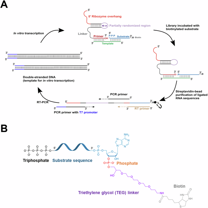
A An RNA library containing a partially randomized sequence having an estimated complexity of ~1014 sequences (depicted in light purple) derived from an AIP-Ligase (RS1)12 was challenged with an RNA substrate (depicted in light blue), containing a 5’-triphosphate group and a 3’-TEG-biotin group (depicted in gray) in the presence of an RNA template (depicted in green). Ligated sequences were purified by binding to streptavidin-coated magnetic beads and reverse transcribed using a primer (RT primer; depicted in gold) that is complementary to the entire substrate sequence. The RT primer also has the potential to bind directly to the library sequences by forming four base-pairs with the 3’ end of their ‘primer’ sequence (depicted in red). The cDNA was PCR-amplified, with the T7 promoter sequence (depicted in blue) added to the dsDNA sequence during PCR. This dsDNA was transcribed to generate the library for subsequent rounds of selection. All sequences are included in Supplementary Data Table 1. B The chemical features of the substrate used in the selection (PPP-Substrate-Biot). The substrate contains a triphosphate group (black) on its 5’ end and is connected to a biotin moiety (gray) via a triethylene glycol (TEG) linker (purple) attached to the 3’-phosphate (orange) on the terminal adenine of the substrate.
Outputs from each round were analyzed by high-throughput sequencing13. Closely-related sequences isolated from the last round (round 6) were binned into clusters. Collectively, these sequences represented >90% of the entire selected population (Table 1). The peak sequences of the five most abundant clusters, referred to henceforth as CS1-CS5 (Table 1, Supplementary Fig. 1) were 10–28 mutations from the parent AIP-Ligase, RS1 (Table 1). We tested CS1-CS5 for their capacity to ligate to the 5′-triphosphorylated, 3′-biotinylated substrate (henceforth, PPP-Substrate-Biot) used in each selection round (Fig. 1B). CS1, CS2, CS4, and CS5 catalyzed ligation with rates between 0.4 h−1 and 1.5 h−1, yielding between ~20% and ~50% ligated product after 3 h. In contrast, CS3 showed reduced activity with a ~ 75-fold lower ligation rate than CS1 (Fig. 2A, C, D). Interestingly, only CS3 catalyzed the desired ligation reaction with a PPP-Substrate, which we previously reported as a bona fide triphosphate ligase ribozyme11. CS1, CS2, CS4, and CS5, on the other hand, appeared to exhibit an unexpected dependence on the 3′ biotinylation state of the substrate (Fig. 2B). This indicated that CS1, CS2, CS4, and CS5 do not catalyze the desired reaction. However, the appearance of a ligated product when incubated with PPP-Substrate-Biot suggests that these sequences are ligases that use an unforeseen reaction pathway. In the following sections, we investigate the unexpected features of these ligases and uncover a novel enzyme reactivity.
Fig. 2: Ligase activities of the isolated sequences.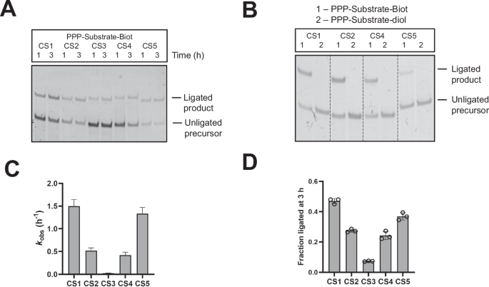
A Peak sequences from clusters 1–5, CS1-CS5, catalyze ligation with a substrate containing a 5’-triphosphate group and 3’-TEG-biotin group (PPP-Substrate-Biot). B CS1, CS2, CS4, and CS5 do not ligate to a substrate oligonucleotide with a 2’,3’ cis-diol (PPP-Substrate-diol). Ligation was assayed at 3 h. C CS1, CS2, CS4, and CS5 exhibit kobs values of 0.4–1.5 h-1, but CS3-catalyzed ligation is significantly slower with PPP-Substrate-Biot. Error bars indicate standard error of the mean (S.E.M). D CS1, CS2, CS4, and CS5 ligated to 20–50%, while CS3 ligated to ~7% with PPP-Substrate-Biot in 3 h. Error bars indicate standard deviation. Data in (C) and (D) were obtained from triplicate measurements. Ligation reactions contained 1 µM ribozyme, 1.2 µM RNA template, and 2 µM RNA substrate, PPP-Substrate-Biot, in 100 mM Tris-HCl (pH 8.0), 300 mM NaCl, and 100 mM MgCl2. Experiments were performed at least in triplicate. Source data are provided as a Source Data file.
Table 1 Sequence alignment of the isolated ligase ribozymesUnexpected reaction between the substrate 2′-hydroxyl and the ribozyme 5′-triphosphate groups
Given that CS1, CS2, CS4, and CS5 all exhibited an identical apparent dependence on the 3′ biotinylation state of the substrate, we selected the most abundant and active sequence, CS1, for detailed biochemical characterization. The desired reaction was a templated ligation between the 3′ and 5′ termini of the ribozyme and substrate, respectively, where the 16-nt template oligonucleotide was expected to bring the two RNA termini together by forming 8 base-pairs with sequences at the 3′ and 5′ end of each RNA (Fig. 1A). However, CS1-catalyzed ligation was only 3-fold slower in the absence of the external template (Fig. 3A, B). This absence of template requirement was reminiscent of a previous in vitro selection experiment that unintentionally isolated ribozymes that catalyze a ligation reaction between the ribozyme 5′-PPP and the 5′-phosphorimidazole moiety of an RNA substrate, generating a 5′-5′ linkage9. To test the possibility that we had accidentally selected similar 5′-5′ ligases, we assayed the ligase activities of truncated versions of CS1, created by deleting either its first 25 nucleotides (5′ truncation or 5′t) or its last 14 nucleotides (3′ truncation or 3′t). While the 3′ truncation preserved ligation, albeit 8-fold slower, the 5′ truncated ribozyme was inactive (Fig. 3C, D). This suggested that the deleted 25-nt sequence at the ribozyme 5′ end is important for its activity, either by being part of its active fold or by directly participating in ligation. To decouple the potential structural vs. functional roles of the 5′ sequence of CS1, we tested CS1 variants containing either a triphosphate (PPP), monophosphate (P), or hydroxyl (OH) group at their 5′ ends against substrate variants with either a 5′-PPP, 5′-P, or 5′-OH. While all substrate variants ligated to CS1, only a 5′-triphosphorylated CS1 retained activity (Fig. 3E). Efficient ligation with 5′-P or 5′-OH substrates discounted the possibility of a nucleophilic attack by the ribozyme 5′-PPP on the substrate 5′ end as observed in the case of the 5′-5′ ligase9. The inactivity of CS1 without a 5′-PPP, on the other hand, indicated that the ribozyme 5′ end was the likely site of nucleophilic attack by the substrate. In principle, a promiscuous ribozyme could catalyze the nucleophilic attack of substrate 5′-PPP, 5′-P, or 5′-OH groups on its own 5′-α-phosphate, with the release of a pyrophosphate group6,14,15. However, similar reactivities of all three substrates, under a range of different conditions, suggested that the 5′ end of the substrate did not participate in ligation. CS1 exhibited similar reaction rates (kobs = ~1.5 h−1) with all three 5′-modified substrates (Supplementary Fig. 2) and exhibited comparable Mg2+ requirements with substrates possessing 5′-PPP or 5′-OH groups: [Mg2+]1/2 (OH-Substrate-Biot) = ~15 mM; [Mg2+]1/2 (PPP-Substrate-Biot) = ~20 mM (Supplementary Fig. 3). Additionally, CS1 showed similar pH-rate profiles (log kobs vs pH) when ligating 5′-P or 5′-OH substrates, exhibiting linearity with a slope of ~1 between pH 6.5 and 8 for both reactions (Supplementary Fig. 4). This usually indicates a single H+ transfer in the rate-determining step in this pH regime involving the nucleophilic hydroxyl group on the substrate16. The irrelevance of the substrate 5′ chemistry was conclusively shown by the ligation of CS1 to a substrate variant containing an inverted dideoxythymidine(ddT) group blocking its 5′ end (Fig. 3F).
Fig. 3: Untemplated ligation between the 5’-triphosphate group of CS1 and the 2’-hydroxyl group of the substrate.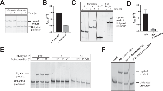
A, B CS1-catalyzed ligation is preserved in the absence of an external template with only a 3-fold decrease in activity. C Truncating CS1 by deleting its first 25 nucleotides abrogates ligation; however, deleting its last 14 nucleotides preserves activity. D Deleting 14 nucleotides from the 3’ end of CS1 results in 8-fold slower ligation than ligation catalyzed by a full-length CS1 ribozyme. Data in (B) and (D) were obtained from triplicate measurements, where error bars indicate standard error of the mean (S.E.M). E Ligation requires a triphosphate group at the ribozyme 5’ end but is agnostic to the chemistry at the substrate 5’ end. F Blocking the 5’ end of the substrate with an inverted dideoxythymidine group (ddT-P-Substrate-Biot) preserves ligation; however, a substrate with a 2’ terminal deoxyribonucleotide (P-Substrate16dA-Biot) fails to ligate to CS1. Ligation reactions contained 1 µM ribozyme and 2 µM RNA substrate (PPP-Substrate-Biot unless otherwise specified) in 100 mM Tris-HCl (pH 8.0), 300 mM NaCl, and 100 mM MgCl2. Reactions do not contain an external template unless indicated (A) at 1.2 µM concentration. Ligation reactions in (E) and (F) were assayed at 3 h. Experiments were performed at least in triplicate. Source data are provided as a Source Data file.
With the 5′ end of the substrate discounted as a reactive center and the 3′ end of the substrate being blocked by TEG-biotin, we turned to the 2′-OH of the terminal adenine residue of the substrate as the most likely candidate for the nucleophile. A substrate where the terminal adenine ribonucleotide (rA16) was replaced by a deoxyribonucleotide (dA16) was inactive for ligation, confirming the substrate terminal 2′-OH as the nucleophile (Fig. 3F and Supplementary Fig. 5A). Identical results with CS2, CS4, and CS5 established a common reactivity for all four ligases (Supplementary Fig. 5B). Nucleophilic attack of the substrate 2′-OH on the ribozyme 5′-PPP would generate a noncanonical 2′-5′ linkage between the ribozyme and substrate. We performed a dual exonuclease digestion assay to test for the presence of this noncanonical 2′-5′ linkage. We purified the products of ligation between CS1 and substrates with either 5′-PPP or 5′-P groups and subjected them to 5′-P-dependent Terminator 5′→3′ exonuclease digestion. As expected, the 5′-PPP ligated product was resistant to degradation (negative control), but the digestion of the 5′-P ligated product yielded RNA that was comparable in size to the ribozyme (Supplementary Fig. 6A). On the other hand, 3′→5′ RNase R digestion of the ligated product generated RNA that was one nucleotide longer than the substrate (Supplementary Fig. 6B). These results suggest the presence of a 2′-5′ phosphodiester bond between the ribozyme and substrate and are consistent with a reaction between substrate terminal 2′-OH and ribozyme 5′-PPP groups.
The reactivity described above also explains the higher-order ligation products generated during CS1-catalyzed ligation with a 5′ 2AI-activated substrate (AIP-Substrate-Biot) in the presence of the external template and 100 mM Mg2+ (Supplementary Fig. 7). These higher-order products are concatemeric RNA sequences with a heterogenous backbone composed of both 2′-5′ and 3′-5′ phosphodiester linkages formed as a result of a combination of ribozyme-catalyzed 2′-5′ ligation and template-directed nonenzymatic 3′-5′ ligation at a high Mg2+ concentration. The production of concatemeric RNA demonstrates the ribozyme′s ability to function in the context of longer transcripts.
Ligation requires a 3′-phosphate group on the substrate
The unexpected observation that substrates without the 3′-‘TEG-biotin′ moiety (i.e., with a 2′,3′ diol) are not substrates for ligation catalyzed by CS1, CS2, CS4, and CS5 (Fig. 2B) pointed to a potential role for the 3′-biotin tag in this reaction. As ribozymes that utilize thiamin (Vitamin B1) as cofactor have been reported17, we wondered if CS1, CS2, CS4, and CS5 might utilize biotin (Vitamin B7) for catalysis. We found that supplementing a reaction between CS1 and an unbiotinylated substrate with free biotin did not rescue ligation (Fig. 4A), moving the biotin modification from the 3′ to the 5′ end of the substrate eliminated ligation (Fig. 4A), and replacing the biotin moiety with desthiobiotin (biotin without a S atom) preserved ligation (Figs. 1B, 4B). These results collectively cast doubt on the direct involvement of biotin. Deleting the TEG spacer between the substrate 3′-phosphate and the biotin (so that the 3′-phosphate was directly connected to the biotin) also preserved ligation (Fig. 1B, Fig. 4B). Importantly, CS1 ligated a substrate that lacked TEG-biotin but possessed a 3′-P group (Substrate-3′P) with rates comparable to that of a biotinylated substrate (Fig. 4B, D). Therefore, the apparent dependence of CS1 on the substrate 3′-‘TEG-biotin′ was, in fact, a requirement for a 3′-phosphate group on the substrate. CS2, CS4, and CS5, like CS1, ligated to Substrate-3′P revealing that the isolated ribozymes represented a new class of ligases that catalyze a reaction between its 5′-triphosphorylated end and the 2′-OH group of a 3′-phosphorylated oligoribonucleotide substrate (Fig. 4D, E).
Fig. 4: Ligation requires a 3’-phosphate group on the substrate.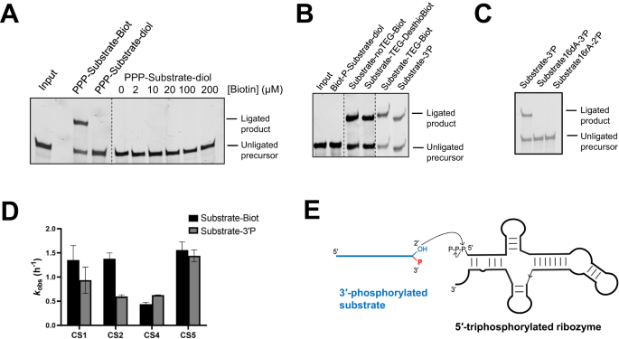
A Ligation with an unbiotinylated substrate containing a terminal cis-diol is not rescued upon the addition of free biotin. B Ligation is abolished when the TEG-biotin moiety is moved to 5’ end of the substrate, but a substrate with the biotin group replaced by a desthiobiotin group retains the ability to be ligated. A substrate with a 3’-phosphate group is active for ligation. C A substrate with 2’ phosphate and 3’ hydroxyl groups (Substrate16rA-2’P) is not ligated, indicating that only the 2’-OH is catalytically-activated for nucleophilic attack in the presence of a vicinal 3’-P, but not a 3’-OH in the presence of a vicinal 2’-P. D Ligation rates for CS1, CS2, CS4, and CS5 with substrates containing terminal TEG-biotin or 3’-phosphate groups are comparable. Data were obtained from triplicate measurements, where error bars indicate standard error of the mean (S.E.M). E The isolated ligase activity involves the nucleophilic attack of the 2’-OH group of a 3’-phosphorylated substrate on the 5’-triphosphate group of the ribozyme. The secondary structure of the ribozyme depicted here is the SHAPE-derived structure of CS1 (see Supplementary Fig. 13B). Ligation reactions contained 1 µM ribozyme, 1.2 µM template and 2 µM RNA substrate in 100 mM Tris-HCl (pH 8.0), 300 mM NaCl, and 100 mM MgCl2. Biotin was added to the reaction as indicated in (A). Ligation reactions in (A–C) were assayed at 3 h. Experiments were performed at least in triplicate. Source data are provided as a Source Data file.
The essential role for the substrate 3′-phosphate is underscored by the rate acceleration of over 5 orders of magnitude relative to the background ligation rate (background ligation between a 5′-PPP-RNA oligonucleotide corresponding to the first 25 nt of the ribozymes and a 5′-FAM labeled 3′-phosphorylated substrate was measured as 2.4 × 10⁻⁶ h−1 at pH 8 and 100 mM Mg2+). Although a 3′-P can, in principle, accelerate substrate ligation by activating the vicinal 2′-OH nucleophile by acting as a general base catalyst, similar ligation rates with Substrate-3′P, which contains a phosphomonoester with a pKa ∼7 and Substrate-TEG-Biot (Fig. 4D), which contains a phosphodiester with a pKa ∼2, discount this potential catalytic role. In fact, the presence of 3′-phosphate or 3′-phosphodiester groups lowers the nucleophilicity of the vicinal 2′-hydroxyl group18, which is supported by a slight increase in the 2′-OH pKa in the presence of a 3′-phosphate relative to a 3′-hydroxyl (~13.4 vs. ~12.4)19. A more likely role of the 3′-P is in localizing catalytic divalent cations to the active site. Ribozymes use active site-bound divalent metal ions in diverse catalytic mechanisms, including RNA cleavage and RNA ligation reactions20,21,22. Crystal structures of the L1 ligase and the class I ligase ribozymes, both of which catalyze ligation between the substrate 3′-OH and a ribozyme 5′-PPP, reveal that an active site Mg2+ interacts with a non-bridging oxygen on the α-phosphate of the ribozyme 5′-PPP and also with the ribozyme phosphodiester backbone23,24. In addition, the structure of the class I ligase suggests a possible interaction between Mg2+ and the 3′-O nucleophile24. Similar interactions in these ligases between active site Mg2+ ions and the 3′-P group of the substrate and pyrophosphate leaving group of the ribozyme 5′-PPP could be important in catalysis. To examine the nature of the interaction between the terminal phosphate group and Mg2+, we measured reaction kinetics with substrates containing either a terminal phosphate or a thiophosphate in the presence or absence of the thiophilic cation Cd2+ in the background of Mg2+. We observed a 2-fold reduction in ligation rate with a terminal thiophosphate-containing substrate (Substrate-3′sP). When the reaction was supplemented with Cd2+ (25 µM or 1 mM), ligation was restored with a metal rescue value of ~2.5 (Supplementary Fig. 8A, B). More pronounced thio effects and metal rescue values have been reported in ribozymes where Mg2+ directly interacts with either of the non-bridging oxygens of the phosphate group22. A diminished effect is expected as our experiment does not involve a stereospecific O to S substitution in the phosphate. The modest effect we observed might also reflect a weak inner-sphere Mg2+ interaction. Substitution of Mg2+ with the divalent metal ions Ba2+, Ca2+, Co2+, Cu2+, Mn2+, Ni2+ abolished ligation, consistent with a possible catalytic role for Mg2+ (Supplementary Fig. 8C). The specific requirement for the substrate terminal configuration, i.e., 2′-OH, 3′-P was further highlighted by the lack of reactivity of a substrate with a 3′-OH, 2′-P terminus (Substrate16rA-2′P) (Fig. 4C).
As the isolated ribozymes catalyze substrate ligation to their 5′ ends as opposed to their 3′ ends, we were surprised that these sequences could be reverse transcribed at 42 °C using a primer (RT primer) that binds to its 3′ end by forming just 4 base-pairs. This indicates that the isolated sequences survived this step in the selection cycle by exploiting weak interactions between the RT primer and their 3′ ends (Supplementary Fig. 9). Because this ligation pathway joins the 2′ end of the substrate to the 5′ end of the ribozyme, the external template cannot be responsible for bringing these two ends into close proximity, as it does for the expected ligation junction between the ribozyme 3′ end and the substrate 5′ end (Supplementary Fig. 9). Furthermore, since the CS1 ribozyme does not require the external template, we were surprised to find that CS2, CS4, and CS5 did require an external template for activity (Supplementary Fig. 10A). We hypothesized that this template dependence was due to functional base-pairing interactions between the ribozyme 3′ sequence (‘primer′) and the template (Supplementary Fig. 10B). Disrupting base pairs between the primer and the template through template mutations eliminated ligation, which was rescued by compensatory mutations in the ribozyme 3′ primer, supporting this hypothesis (Supplementary Fig. 10C). We found similar interaction between the 3′ end of CS1 and the template (Supplementary Fig. 10D), which explains the decrease in ligation observed in the absence of the template (Fig. 3A, B) and the 3′-truncated version of this ribozyme (Fig. 3C, D). Base-pairing interactions between the template and both the 3′ end of the ribozyme and the 5′ end of the substrate may make this three-component reaction pseudo-intramolecular, thereby stimulating ligation.
Comparative structural analysis of the isolated ligase ribozymes
Although at first glance, CS1, CS2, CS4, and CS5 diverge from each other by 11–22 mutations (Supplementary Fig. 11), which could indicate distinct structural folds, upon more careful inspection, we found that the sequences, especially CS1, CS2, and CS4, could be aligned to highlight structural similarities (Supplementary Fig. 12). To get a better understanding of their secondary structures, we performed SHAPE-probing on CS1, CS2, CS4, and CS5. Constraining their computationally predicted secondary structure with SHAPE reactivities yielded structures that differed significantly from each other and from their parent ligase, RS1 (Supplementary Fig. 13). Despite this apparent structural divergence, we noticed similarities in SHAPE reactivity patterns across all four sequences (Supplementary Fig. 12). The combination of common SHAPE reactivity patterns and comparative sequence analysis revealed the possibility that these ligases, at least CS1, CS2, and CS4, share a common fold.
We identified regions that may form conserved base-paired stems in all four RNAs (Supplementary Fig. 12). In CS1, CS2, and CS4, one of the putative stems consists of 5′-CCACUCA-3′ and 3′-GGUGAGU-5′ regions, while in CS5, the putative stem is shorter, composed of 5′-CUCA-3′ and 3′-GAGU-5′ (shown in green in Supplementary Fig. 12). The presence of this stem in all four RNAs is supported by their overall insensitivity to SHAPE modification. The SHAPE-derived structures of CS1, CS2, and CS4 feature this stem, whereas in CS5, the relevant residues (5′-CUCA-3′ and 3′-GAGU-5′) appear unpaired despite showing low reactivities (Supplementary Fig. 13). The fact that this stem is composed of nucleotides in the constant region and those that emerged as a result of selection increases the likelihood of its presence in these ribozymes. The presence of a second putative stem composed of 5′-GGACAGCG-3′ and 3′-CCUGUCGC-5′ regions (shown in blue in Supplementary Fig. 12) is less certain as residues in the 3′-strand sequence were reactive in SHAPE experiments in CS1 and CS4, indicating a lack of base-pairing. Further prediction is complicated by the lack of reactivity data for a significant portion of this region (Supplementary Figs. 12, 13). Even without SHAPE data for the 3′ end of the ribozymes, the fact that the last eight nucleotides of CS1, CS2, CS4, and CS5 base-pair with the template during ligation (Supplementary Fig. 10) indicates that they are likely unpaired in the ribozyme′s secondary structure. We also noticed high SHAPE reactivities in the 5′-AAUGA-3′ region (residues 66-70) in all four ribozymes, indicating that this region is unpaired, further pointing to the possibility of a shared structure.
In light of these observations, we queried for a common secondary structure for CS1, CS2, CS4, and CS5 using TurboFold, an iterative probabilistic RNA secondary structure prediction algorithm for estimating common secondary structures for multiple sequences, even when they exhibit substantial divergence25. TurboFold outputs converged on a common secondary structure for CS1, CS2, and CS4, but predicted a different structure for CS5 (Supplementary Fig. 14). Structures of CS1, CS2, and CS4, as predicted by TurboFold, featured a base-paired stem composed of 5′-CCACUCA-3′ and 3′-GGUGAGU-5′ regions similar to their SHAPE-derived structures; however, unlike its SHAPE-derived structure, the structure of CS5 obtained from TurboFold featured the shorter stem composed of 5′-CUCA-3′ and 3′-GAGU-5′, we identified from sequence analysis (highlighted by a green a box in Supplementary Fig. 14). TurboFold did not predict the existence of the second putative stem composed of 5′-GGACAGCG-3′ and 3′-CCUGUCGC-5′, which is consistent with the SHAPE reactivity of certain residues in this region. Some of the inconsistencies between the secondary structures derived from SHAPE probing and those predicted by sequence inspection or TurboFold may arise due to misfolding of the ribozyme in the absence of the template and substrate RNAs in SHAPE experiments. Regardless, in the absence of high-resolution structures, it is difficult to claim structural relationships between these ligases, although there are likely significant similarities due to the observations outlined above.
Specific capture and amplification of 3′-phosphorylated RNA
This reactivity of ligating specifically to 3′-phosphorylated RNAs exhibited by the isolated ribozymes is also interesting in the context of extant biology. RNAs with terminal phosphates in the form of 2′, 3′-cyclic phosphates (cP) and 3′-phosphates (3′-P) are generated as products of enzymatic cleavage pathways in RNA processing and maturation and have been implicated in diseases including cancers, amyotrophic lateral sclerosis, tuberculosis, and Parkinson′s disease26,27,28,29,30,31. However, cleaved RNAs constitute a poorly characterized portion of the transcriptome, primarily because they remain invisible to standard library preparation protocols used in high-throughput sequencing. This is due to the inability of 3′-phosphorylated cleaved RNAs to ligate to the 3′ sequencing adapter because they lack free 3′-hydroxyl groups. The handful of recently reported methods for sequencing terminal phosphate-containing RNAs cannot distinguish between cP and 3′-P-containing RNAs due to the promiscuity of the RNA ligases used (Arabidopsis thaliana tRNA ligase or RtcB ligase) or rely on indirect enrichment of cP-RNAs via periodate cleavage of RNA terminal diols32,33,34,35. As the ribozymes identified in this work show absolute discrimination between 3′-OH and 3′-P RNA termini we explored their potential application as reagents for the specific enrichment of 3′-phosphorylated RNA from total cellular RNA.
We found the CS1 has a low KM value of 0.1649 ± 0.042 µM and a high catalytic efficiency (kcat/ KM value) of 27.65 µM−1 h−1 (7670 M−1 s−1) indicating that CS1 could function as a potential reagent for enriching 3′-P-RNAs (Supplementary Fig. 15). We spiked in different concentrations a 5′ FAM-labeled 3′-phosphorylated substrate (FAM-Target RNA-3′P) into an E. coli-derived tRNA mix and incubated this mixture with CS1. The appearance of a band corresponding to the ligated product even when tRNAs were in 60-fold excess supports the ribozyme′s ability to enrich 3′-phosphorylated RNAs from a heterogeneous mixture of cellular RNA (Fig. 5A). The fluorescence signal from the ligated product increased linearly with substrate concentration between 0.05 µM to 0.4 µM, suggesting a potential for quantitative detection of 3′-P-RNAs (Fig. 5B). The captured 3′-P-RNAs must be first reverse transcribed and then PCR-amplified to generate material that can be sequenced (Supplementary Fig. 16A). To see whether the 3′-P next to a 2′-5′ phosphodiester linkage would prevent reverse transcription, we compared RT-PCR before and after removing the phosphate group by shrimp alkaline phosphatase (SAP). Interestingly, the target RNA was amplified even without SAP treatment, showing that reverse transcriptase can copy across the unusual 2′-5′ phosphodiester linkage harboring an adjacent 3′-phosphate (Supplementary Fig. 16B).
Fig. 5: Ribozyme-assisted capture of 3’-phosphorylated RNA.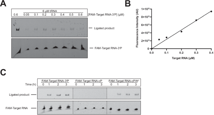
A CS1 captures a FAM-labeled 3’-phosphorylated substrate, FAM-Target RNA-3’P, from a heterogeneous mixture of cellular tRNAs. B Capture, as measured by the fluorescence intensity of the ligated product, is linear in response to substrate concentration between 0.05 µM and 0.4 µM. C CS1 shows specificity toward substrate 3’ termini. It specifically ligates to substrates with 3’-phosphate groups, while being inert to those terminating in 2’, 3’-cyclic phosphate (cP) groups. Acid hydrolysis of the terminal cP group makes these RNAs substrates for ribozyme-assisted capture. Ligation reactions contained 1 µM ribozyme, CS1, and the indicated amounts of FAM-Target RNA-3’P (A, B) or 2 µM RNA targets (FAM-Target RNA-3’P or FAM-Target RNA-cP) in 100 mM Tris-HCl (pH 8.0), 300 mM NaCl, and 100 mM MgCl2. Capture reactions in (A) were assayed at 3 h. Experiments were performed in triplicate. Source data are provided as a Source Data file.
These ribozymes may also be used to indirectly capture cP-RNAs by converting their cP ends to 3′-P (and 2′-P) by mild acidification. Upon incubating a cP-containing substrate (FAM-Target RNA-cP) pre-treated with 10 mM HCl (0 °C/3 h)35 with CS1, we observed a steady increase in ligation with increasing incubation time, indicating cP-RNA capture. FAM-Target RNA-cP, not subjected to the acidification step, was not captured by the ribozyme (Fig. 5C). As self-cleaving ribozymes generate cleavage products with cP ends, this ribozyme-assisted enrichment method, in conjunction with standard RNA-seq, may be used in high-throughput screens to discover new self-cleaving ribozymes36,37. For maximal utility as RNA sequencing reagents, ribozymes must enrich RNAs in a sequence-general manner. Therefore, we tested the substrate scope of ribozyme-assisted target capture. We tested CS1 against truncated versions of the Substrate-3′P, which has the sequence: 5′-ACCACCGCAUUCCGCAp-3′, where A contains the 2′-OH nucleophile. CS1 captured a target representing the last 8 residues of Substrate-3′P (5′-AUUCCGCAp-3′) but was unable to ligate an oligoribonucleotide representing its first 8 residues (5′-ACCACCGCp-3′) (Supplementary Fig. 17A). Target 5′-AUUCCGCAp-3′ was further truncated to two 5 nt pieces and tested for ligation. Once again, the piece containing the nucleophilic adenine (5′-CCGCAp-3′) was captured but 5′-AUUCCp-3′ was not (Supplementary Fig. 17A). Shorter oligomers were not tested due to the difficulty in resolving captured products from the ribozyme by denaturing gel electrophoresis. Substrates, where their terminal A was replaced by C or U, could not be captured by CS1, and a substrate with a 3′ terminal G showed ~500-fold slower ligation with CS1 (Supplementary Fig. 17B, C). CS2, CS4, and CS5 showed similar dependence on the identity of the 3′ terminal residue of the substrate (Supplementary Fig. 18). These results indicate the importance of the adenine nucleotide at the 3′ end of the substrate. Similarly, the ribozyme 5′ end was constrained to a guanine; a G to A mutation in all four ribozymes abolished ligation with all four substrate variants (Supplementary Figs. 17D, 18). These constraints on both ligation junction nucleotides suggest that there is an internal template that brings them together to allow ligation; however, the identity of this internal template, if it exists, remains to be defined.