
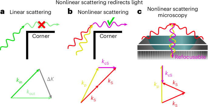
Guided by the theoretical analysis presented above, we implemented an excitation geometry optimized for the direct generation of backward-directed CSRS. The Stokes beam at 1,030 nm was shaped into an azimuthally polarized ring filling the outer annulus of the objective pupil, while the circularly polarized pump beam at approximately 790 nm was confined to the central pupil region.
To enhance the nonlinear signal without increasing the average power delivered to the sample, we reduced the effective repetition rate of both beams to 0.71 MHz using a pulse-picking scheme (Fig. 3a). In addition, boxcar gating of the InGaAs avalanche photodetector reduced the effective dark noise by approximately one order of magnitude. Further details about the experimental implementation are presented within Supplementary Information section 1.
Fig. 3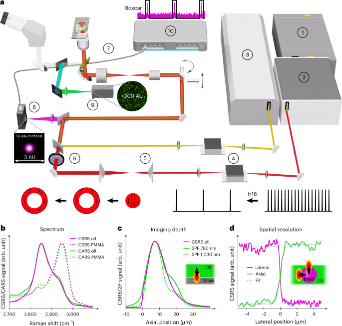
Experimental set-up and characterization. a, Schematic of the experiment: (1) Yb pump laser; (2) amplifier; (3) optical parametric oscillator (OPO); (4) acousto-optic modulator (AOM); (5) double axicon for Stokes ring shape illumination; (6) vortex plate for Stokes azimuthal polarization; (7) laser-scanning microscope (LSM); (8) InGaAs avalanche photodiode (APD); (9) photomultiplier tube (PMT); (10) lock-in amplifier. The PMT features a much larger detection area than an Airy disk (1 Airy unit, AU), while the APD provides a quasi-confocal detection of the CSRS signal. b, CSRS spectra of oil and PMMA obtained by tuning the pump wavelength. c, Depth profiles of CSRS in oil and two-photon fluorescence (2PF) of yellow marker dissolved in oil. The depth limitation of backscattering CSRS arises from the aberration corrections of the TIRF objective and similarly affects two-photon fluorescence. d, CSRS lateral profile extracted from a PMMA–sunflower-oil interface. The error-function fit to the edge-spread function yields an effective lateral line-spread-function FWHM of approximately 330 nm. Axial edge measurements across the same type of interface yielded an effective axial line-spread-function FWHM of approximately 0.92 μm.
As a first quantitative benchmark, we compared the signal strength of forward CARS with backward CSRS. Detailed photon-flux, detector-noise and signal-to-noise ratio calculations are provided in Supplementary Information section 3. Under excitation conditions identical to those used in the simulations and calibrated using detector specifications, we estimate a directly backscattered CSRS photon flux of approximately 7 × 10⁹ photons s−1 emitted from the focal volume in sunflower oil. Switching from purely linear excitation to azimuthally polarized Stokes illumination combined with a circular polarized pump increased the CSRS signal by about 50%, consistent with the predicted polarization dependence.
To rule out any contribution from forward-generated CSRS reflected into the detection path, we placed a mirror directly above the sample. This produced no measurable increase in the detected CSRS photon flux, supporting an intrinsically backward-emitted origin. Any residual forward CSRS under annular Stokes illumination is further suppressed by the small effective collection area of the avalanche photodetector (~3 Airy units), which acts as a confocal pinhole.
For comparison, forward-generated CARS photons can be detected in the backward direction when a mirror is placed above the sample, yielding photon fluxes of approximately 4 × 10¹⁰ photons s−1 (Supplementary Information section 3). In realistic imaging scenarios involving thick or heterogeneous samples, however, epi-detected CARS signals arise either from high spatial frequencies at refractive-index interfaces or from forward-generated CARS that is multiply scattered and back-reflected4,9. Such back-reflected CARS cannot be refocused onto the detector plane, precluding efficient fibre coupling, amplification, spectral analysis or accurate point-spread-function characterization. By contrast, backward CSRS is generated directly into the backward hemisphere and remains refocusable, enabling true confocal detection. For the experimentally used excitation geometry, our simulations show that the backward CSRS field refocuses after the objective–tube-lens relay into a compact, near-Gaussian intensity distribution, supporting fibre- or spectrometer-based collection (Supplementary Fig. 5).
Figure 4a shows backscattered CSRS images of polymethyl methacrylate (PMMA) beads dispersed in sunflower oil. The corresponding spectra (Fig. 3b) confirm that, in the absence of electronic resonances, CARS and CSRS provide nearly interchangeable vibrational information10,11. Notably, strong CSRS signals are obtained from homogeneous regions of sunflower oil, well away from any bead–oil or oil–substrate interfaces. In contrast to epi-CARS, the PMMA beads do not exhibit interface-related shadowing or highlighting effects, consistent with the absence of high-pass filtering in backward CSRS.
Fig. 4: Experimental comparison of epi-CARS and backscattered CSRS imaging.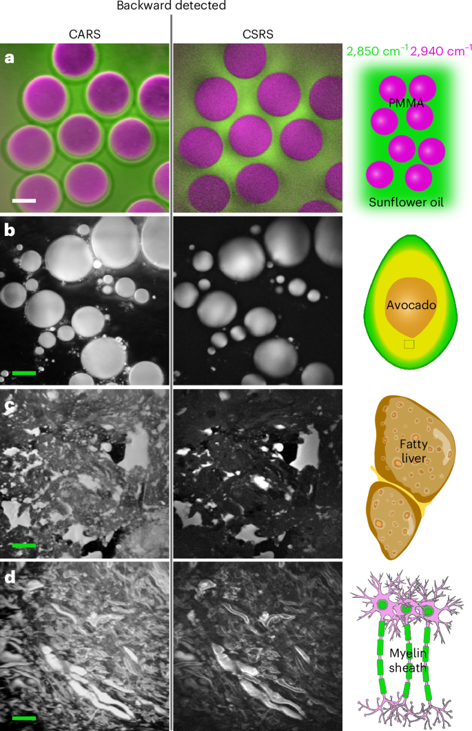
a, Twenty-micrometre PMMA beads dispersed in sunflower oil; green and purple correspond to Raman resonances at 2,850 cm−1 and 2,940 cm−1, respectively. b–d, Avocado flesh (b), human fatty liver tissue (c) and myelin sheaths in sheep brain tissue (d), all imaged at 2,850 cm−1. All scale bars, 10 μm. Effective pixel dwell time: 20 μs. Image acquisition time: 21 s for a 1,024 × 1,024-pixel image. Pump and Stokes beam powers at the sample were 2.5 mW and 6 mW, respectively.
To assess a possible contribution of high spatial frequencies at the coverslip–oil interface to the detected CSRS signal, we investigated the axial signal dependence using a total internal reflection fluorescence (TIRF) objective (Fig. 3c). Such objectives are designed to form a diffraction-limited focus at the coverslip interface, but the focus quality degrades rapidly with increasing depth12. When imaging deeper into the sample, it is therefore essential to distinguish between limitations imposed by CSRS phase matching and those arising from point-spread-function degradation.
We further quantified the spatial response from edge measurements at PMMA–oil interfaces. The lateral edge response yields an effective lateral line-spread-function full width at half maximum (FWHM) of approximately 330 nm (Fig. 3d). In addition, axial edge profiles acquired across the PMMA–oil interface yielded an effective axial line-spread-function FWHM of approximately 0.92 μm. These values should be interpreted as edge-derived effective imaging resolutions rather than as a direct measurement of the local nonlinear source distribution alone. In particular, the axial edge response includes coherent CSRS generation, backward propagation, collection through the high-NA objective, and confocal detection.
Having established backward CSRS on well-defined test samples, we next assessed its performance on a biologically relevant yet structurally simple specimen. Avocado flesh provides an intermediate system in which lipid droplets introduce pronounced refractive-index variations while maintaining a comparatively simple microstructure. Figure 4b compares backward CSRS with epi-CARS at the lipid resonance at 2,850 cm−1. Backward CSRS remains robust despite refractive-index mismatches between lipid droplets and the surrounding tissue, which effectively reduce the illumination NA13. Moreover, a fluorescence background is observed in the epi-CARS channel14—even when using a narrowband filter—whereas no such contribution is detected in the CSRS channel at 1,470 nm (Supplementary Fig. 1 and Supplementary Information section 1).
We finally applied backward CSRS to more demanding biological specimens characterized by strong scattering and fine-scale organization. Figure 4c shows backward CSRS imaging of human fatty liver tissue at the lipid resonance at 2,850 cm−1. Lipid droplets and surrounding tissue can be clearly distinguished. Fatty liver disease is clinically highly relevant, as hepatic lipid accumulation constitutes an early and potentially reversible stage of metabolic liver disorders15. Notably, the brightness difference between lipid-rich regions and surrounding tissue is reduced in epi-CARS compared with backward CSRS. This difference is consistent with the distinct spatial transfer functions of the two epi-detected signals. In thick tissue, epi-CARS generally contains a mixture of true backward-emitted CARS generated by high-spatial-frequency structures near the focus and originally forward-emitted CARS that is subsequently scattered or reflected back toward the objective. Consequently, epi-CARS images can be influenced by interfaces, local scattering, sample reflectivity and return-path efficiency. This behaviour has been observed directly in spinal cord tissue, where forward- and epi-detected CARS provide related but visibly different myelin contrast and line profiles16. By contrast, backward CSRS is generated directly into the backward hemisphere by nonlinear phase matching and therefore provides bulk chemical contrast without relying on high-pass spatial filtering or scattering-mediated return.
Figure 4d highlights backward CSRS imaging of brain tissue at the lipid resonance at 2,850 cm−1. The bright ellipsoidal structures correspond to myelin sheaths, which are lipid-rich and provide electrical insulation along neuronal pathways. Damage to these structures underlies several neurological disorders, including multiple sclerosis17 and Guillain–Barré syndrome18. Efficient, label-free imaging of myelin therefore represents a key capability for advancing research into demyelinating diseases.