
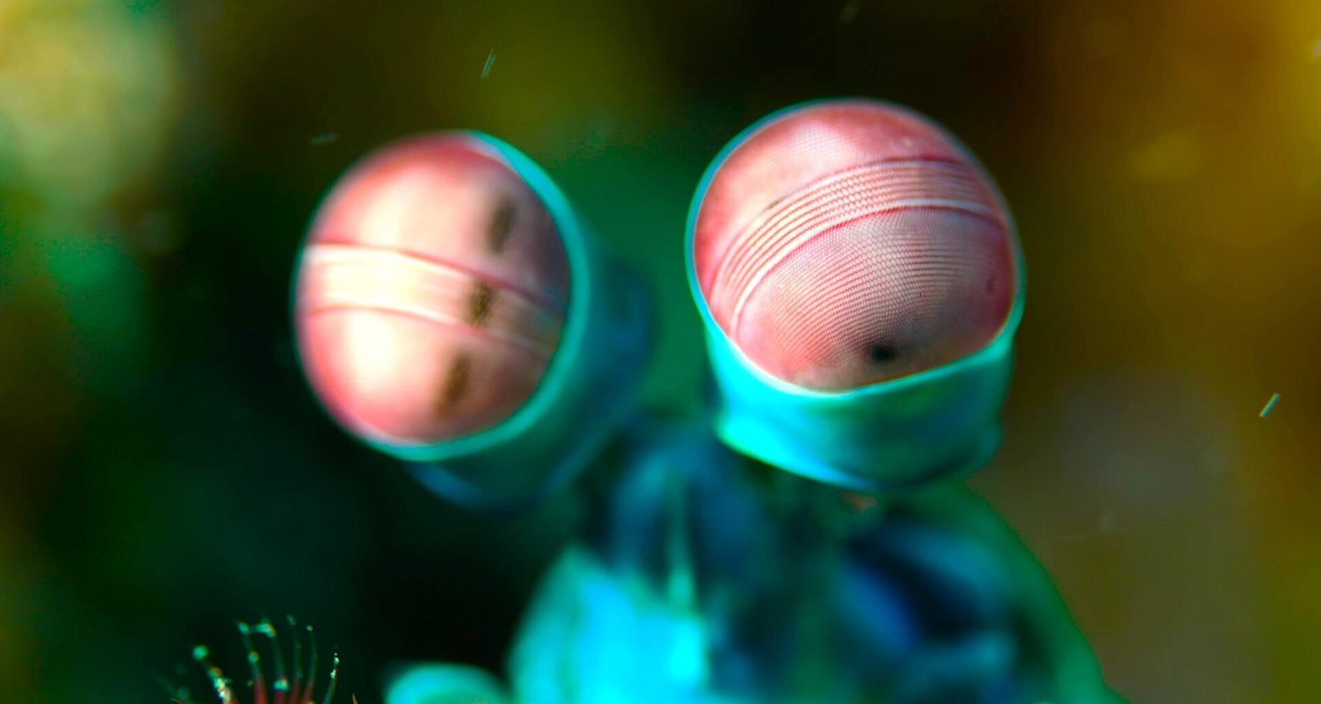
Researchers identified unusual visual pigments that respond to deep red light and can be switched on and off using different colors.
Mantis shrimp have a reputation as the animals that see colors no human ever will.
But new research reveals a surprise. Most of their visual pigments detect green light, while a handful have extraordinary abilities.
Three catch deep red light that almost no other animal can, and four switch on with one color of light and off with another.
By testing the pigments individually for the first time, researchers uncovered how mantis shrimp eyes may detect wavelengths of red light that most animals cannot.
The study was led by Camille Brouillon, Alina Pushkarev, and Marjorie Liénard, researchers affiliated with Humboldt University of Berlin and the GIGA Institute at the University of Liège in Belgium.
None of this was measured in a living mantis shrimp eye. The team built each pigment inside human kidney cells grown in a dish.
There it could be tested on its own, away from the dozens of others packed into the same eye.
Most of the pigments see green
A mantis shrimp eye, the one that earned the animal its name for seeing colors humans can’t, is split into an upper and a lower half. A band of six rows of facets runs between them.
Four of those rows handle color, and each one is stacked in three layers of light-sensing cells.
The pigments live in those cells. Each is a protein called an opsin wrapped around a fragment of vitamin A called retinal, and the retinal is the part that absorbs the light.
Brouillon’s team built 17 of the eye’s opsins and gave each one the same form of retinal that a human eye uses.
Twelve absorbed best near 540 nanometers, which is green, much like the green pigment in a human eye. Two absorbed blue. The last three peaked in the red, at 592 nanometers, 607 and 615.
A red detector is rare in animals. Most pigments built on the ordinary form of retinal stop short of 600 nanometers. Goldfish and salamander pigments reach 617 and 615, but they do it with a modified version of the molecule.
Chemists call the light-catching retinal the chromophore.
“This is the first time such pigments have been described in an animal whilst using the same chromophore as humans,” said Liénard, a molecular biologist at Liège.
Four pigments switch on and off
In human visual pigments, light changes the shape of retinal, triggering a process that requires the pigment to be regenerated.
Four of the shrimp pigments work differently. Light of one color turns them on, light of another turns them off, and the retinal never leaves.
Study senior author Peter Hegemann explained that two of the pigments are activated by red light and deactivated by blue light.
“The other two work the other way round: they are activated by blue light and deactivated by orange light,” Hegemann said.
In the dish, a flash of red light switched one of the red pigments on, and a second flash of blue-green light cut its signal by about three-quarters.
The two blue pigments could be flipped on and off over and over, losing little strength each time. The team calls the four pigments bistable, because each has two stable states.
“Bistable pigments, on the other hand, can be reactivated directly by a second photon of light that restores the retinal, suggesting that the visual system remains continuously operational,” said Brouillon.
Colored filters stretch red vision
In two of the four color rows, a layer of red carotenoid pigment sits between the upper and lower cells, like a tinted lens.
It absorbs the shorter wavelengths before they reach the cells below it. A pigment underneath therefore responds mostly to the red light that gets through.
The team calculated what each filter does to the pigment beneath it. In the upper layer of one row, the two red pigments shift to 632 and 637 nanometers and keep about half of their sensitivity.
In the bottom layer of the same row, the filter is far denser. There, one pigment’s peak moves out to 687 nanometers, deep red near where visible light ends, and it keeps only about 2.5% of its sensitivity.
Earlier estimates for that row, taken from measurements of mantis shrimp eyes themselves, had put its peak at 560 nanometers in this species and 610 in two related ones.
Dusk needs different pigments
A peak at 687 nanometers comes at a price. A dense filter pushes sight deeper into the red, but it absorbs nearly all of the light.
So the far-red cells at the bottom of the stack are probably useful only in bright daylight, the team wrote.
The more sensitive upper cells carry color vision at dawn and dusk, when this species is most active and ventures farthest from its burrow.
“You could compare this to wearing sunglasses during the day and using a torch at dusk,” Liénard said.
“In bright light, when the ultraviolet, blue and green pigments are saturated, the red opsins continue to function. At dawn and dusk, the animal can rely on its dozen green opsins and its bistable blue opsins.”
Why a reef predator wants far-red sight is a separate question. The patches on the club-like front limbs that a mantis shrimp swings at rivals reflect up to 10% of the far-red light that hits them.
The red cells could add a channel for sizing up an opponent, and nobody has tested that in a living animal.
At least 13 of the 17 opsins passed a signal to a G protein, the relay inside the cell that many human hormone and drug receptors also use.
That sets them apart from the algae proteins optogenetics has mostly relied on, which open a pore in the cell rather than triggering a relay.
A pigment that red light turns on and blue light turns off could serve as a reversible switch for those relays, the University of Liège wrote in announcing the work.
Because red light penetrates deeper into tissue and is generally less damaging than blue or ultraviolet light, these pigments could eventually become valuable medical tools.
However, such applications remain experimental.
Mysteries hidden in shrimp eyes
Everything here was measured in cells in a dish, and the team wrote that a pigment can behave slightly differently in a living eye.
Some of the opsins produced no signal at all with the human relay proteins used in the tests. They may pair with a shrimp protein the team did not try.
The findings reveal that mantis shrimp have evolved remarkable ways to detect and control their responses to light.
Scientists are only beginning to understand how these abilities shape the animals’ extraordinary vision – and how they might someday benefit human medicine.
The full study was published in the journal Science Advances.
—–
Like what you read? Subscribe to our newsletter for engaging articles, exclusive content, and the latest updates.
Check us out on EarthSnap, a free app brought to you by Eric Ralls and Earth.com.
—–