
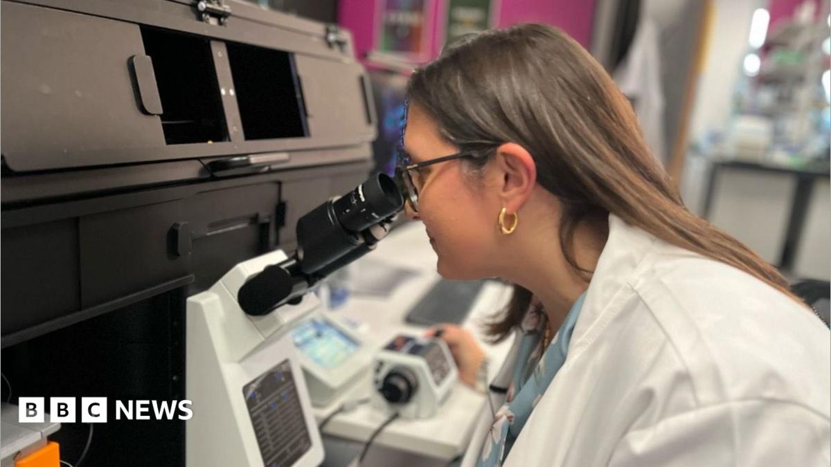
It is hoped a new microscope will help pave the way for a greater understanding of conditions like inflammatory bowel disease.
The Stimulated Emission Depletion (STED) optical microscope will enable researchers at the Rosalind Franklin Institute to study much thicker samples, and in far greater detail.
The scientists who developed the super-resolved fluorescence microscopy technology, used in the STED microscope, won the Nobel Prize for Chemistry in 2014.
Dr Karina Pombo-Garcia, group leader of the lab where the microscope is based, said: “I think it’s going to be a huge game changer.
“It is unique in the UK and one of only a few in world.
“It allows us to look at very, very tiny processes inside cells with a very high resolution and we can look at these processes live.”
The STED microscope uses lasers shaped like a doughnut to vastly increase the resolution of images.
It also allows researchers to label multiple molecules with different coloured fluorescent tags in a single sample.
Dr Pombo-Garcia said that allowed them to look at “how proteins come together to assemble complexes and how these go wrong in disease”.
Unlike other optical microscopes, rather than looking at flat samples on a slide, the microscope allows researchers to study “thick organoids and tissue samples derived from patients”.
“So it helps us to recapitulate better what happens in a real scenario, so that’s very exciting,” she said.
One of the first research projects to use to the microscope is studying how cells form in the creation of the human bowel, and to better understand how those cells then go wrong in conditions like inflammatory bowel disease.