
New studies by two major La Jolla medical research institutions explore aging and its effects on the brain, albeit with different focuses.
Epigenetic atlas
In an effort to better understand the impact of aging on brain function, scientists at the Salk Institute for Biological Studies recently delivered their most comprehensive single-cell atlas detailing epigenetic changes in the brains of mice.
Epigenetic changes are modifications of gene expression that don’t change the underlying DNA sequence.
The new atlas, published March 11 in the scientific journal Cell, details the ways in which genome structure, gene activity and methylation (a biochemical mechanism for regulating gene expression) change across eight brain regions and 36 brain cell types.
“Nearly 900,000 cells were used to trace differences between aging in different brain regions and cell types,” according to the Salk Institute.
The atlas points to epigenetic differences between age groups and forms a basis for developing deep-learning models that can predict further changes, researchers say.
The atlas will serve as a framework for interpreting human brain datasets on Amazon Web Services and the Gene Expression Omnibus. This will include data from the National Institutes of Health’s Brain Research Through Advancing Innovative Neurotechnologies, or BRAIN, initiative.
The project took a total of five years, according to co-author Joseph Ecker, a professor of genetics and director of the Genomic Analysis Laboratory at Salk and an investigator for the Howard Hughes Medical Institute. Four of those years were dedicated to data generation and the last to analysis, he said.
Salk scientists hope the atlas — which is available online — will spur global collaboration.
“Allowing access in different ways is important because you’ve got individual investigators that are looking at individual genes that they want, but others are interested in getting the raw data … and using that for building machine-learning approaches,” said Ecker, who wrote the study with research professor Margarita Behrens.
It also could lay the groundwork for accelerated findings.
“We’re trying to use the mouse brain aging as a model for human brain aging because we can manipulate genes in the mouse brain to test what’s important and not important in aging,” Ecker said.
“In humans, obviously we have genetic differences, we have lifestyle differences, smoking, all sorts of environmental exposures that could alter the brain in ways that could be difficult to study. So now we have features that we can look at and say ‘These are things that are changing, even in a well-controlled mouse study.’”
Newly defined genetic disease
Researchers at Sanford Burnham Prebys and an international team of collaborators explored premature aging and cognitive deficits in a paper released March 19 in Nature Communications.
The study was co-written by Dr. Su-Chun Zhang, a professor and director of the Center for Neurologic Diseases at Sanford Burnham Prebys, and staff scientist Fang Yuan.
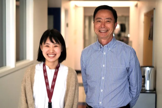 Fang Yuan, a staff scientist at Sanford Burnham Prebys in La Jolla, and Dr. Su-Chun Zhang, director of the institute’s Center for Neurologic Diseases, collaborated on an aging-focused scientific paper released last month. (Sanford Burnham Prebys)
The work traces back several years to when a collaborator of Zhang’s identified a family of patients whose teenage members showed signs of premature aging such as gray hair and eyebrows.
Zhang, then director of the Signature Program in Neuroscience & Behavioral Disorders at Duke-NUS Medical School in Singapore, recalled that “this group of kids not only have premature aging symptoms but also severe neurological problems and cognitive deficits. They do not speak well … and they have shown delayed intellectual capability.”
Zhang and fellow researchers suspected that an unknown disease was at play.
Scientists combined genome sequencing and cellular reprogramming to key in on which gene mutation is at fault for aging and cognitive deficits. They were able to trace it to a mutation in the IVNS1ABP gene, which offers instructional codes to build an influenza virus non-structural protein-1 binding protein.
“Relatively little research has been done on this gene and protein and no one has ever linked them to the biology of aging, premature aging diseases or neuropathy,” Yuan said in a statement.
Among the key findings was that mutation-holding, patient-derived cells grew at a slower rate than a control group reprogrammed from a sibling without the disease, suggesting they had entered a state of senescence, in which aging cells stop dividing but persist, accumulating over time to fuel chronic inflammation, tissue dysfunction and age-related diseases. Further research showed that DNA damage was taking place during cell division.
The results, Zhang believes, emphasize the potential of cellular reprogramming and patient-derived stem cell models in studying rare diseases.
The next step, he said, is to move beyond the current modeling.
“All this is done in a petri dish,” Zhang said. “In a petri dish, you can only do so much; you can only look at so many cell types. You cannot look at the whole body.
“It’s very difficult to ask cells in a petri dish to think or respond. We cannot do it. So that kind of test needs to come from intact animals.” ♦