
Hydroxyapatite is the primary inorganic component of bones and hard tissues, and highly relevant in a variety of medical applications. The incorporation of dopants into the calcium sites of this material introduces atomic defects that endow it with unique luminescent properties for bioimaging applications.
“Density functional theory calculations play a crucial role in understanding the impact of elemental substitution on materials, while allowing for precise control over substitution sites and concentrations,” said Graeve, who is the Elias Masry Endowed Professor in Engineering at the Jacobs School of Engineering Department of Mechanical and Aerospace Engineering. Graeve also serves as director of the Program in Materials Science and Engineering at UC San Diego.
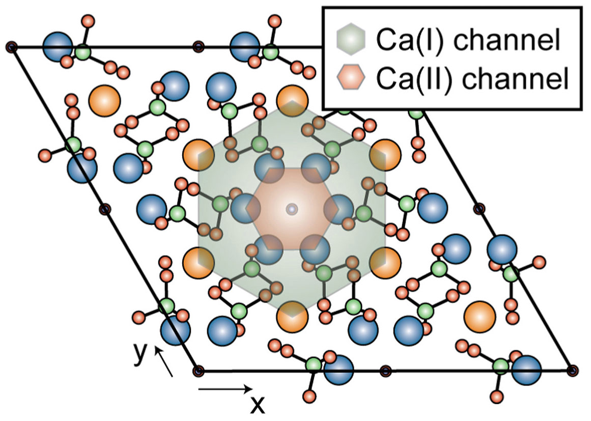
“Our simulations using NSF ACCESS allocations on SDSC’s Expanse showed that hydroxyapatite becomes more structurally stable — meaning its crystal structure is better maintained — when certain hydroxyl groups lose a proton and become oxide ions,” she continued. “That change helps balance europium’s extra charge, making the material more likely to settle into the calcium sites that better support hydroxyapatite’s structure.”
Graeve said that the findings, which have been published in the Journal of Solid State Chemistry, could help scientists design more predictable bone-like materials for future biomedical imaging tools. Because the results also match experimental observations conducted by the team, the study gives researchers more confidence that the computer model reflects real behavior.
The work on Expanse was provided by NSF ACCESS (allocation no. MAT24007).
Read more news about: Halıcıoğlu School of Data Science and Computing,