
Powered by Berkeley News
Who Runs the Negotiation? (Girls)
Human Aging on a Chip
 (Mathew Burciaga/UC Berkeley)
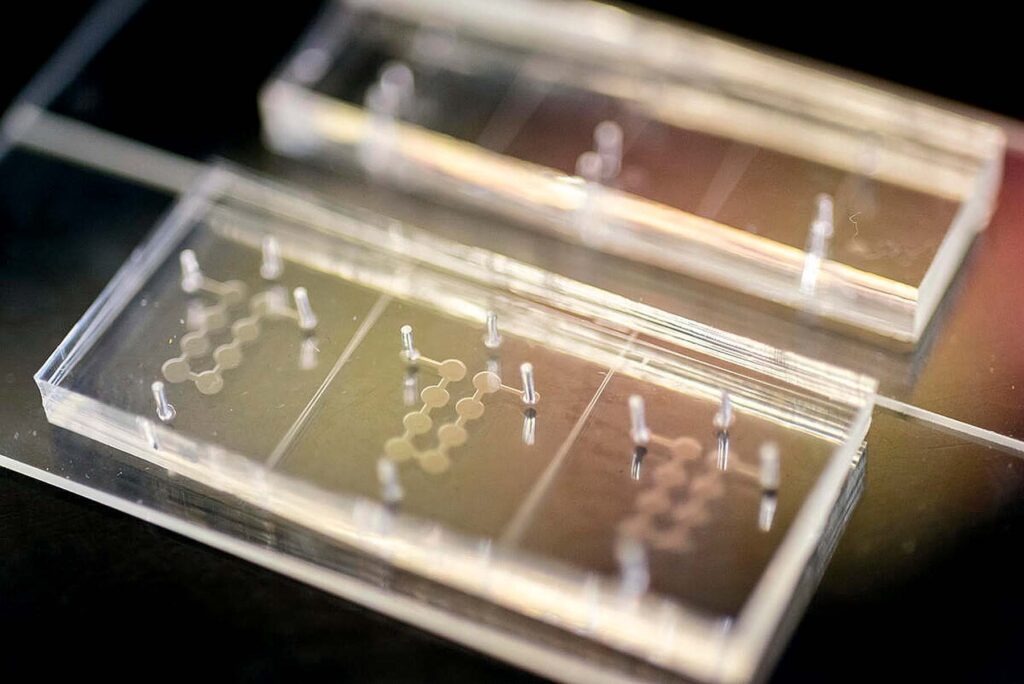
An organ-on-a-chip may sound like science fiction, but scientists have successfully mimicked and miniaturized lungs, the blood-brain barrier, vaginas, and more. Now Berkeley metabolic biologist Andreas Stahl and former longtime Berkeley professor Irina Conboy are adding human aging to that list.
The chip, created at the Biomolecular Nanotechnology Center at QB3, stores fat and liver tissue cells in separate yet connected chambers. Tiny channels stand in for vasculature, which require only a fraction of the fluid typical for standard cell cultures.
The system expands on an earlier one created by Stahl. Conboy, a pioneer in longevity research who has studied aging and reverse aging in mice, wondered if they could use Stahl’s chips to age human tissue.
By exposing the tissue cells in the chip to blood serum from donors aged 62 and older, the researchers were able to induce the telltale signs of aging, including chronic inflammation, astonishingly quickly. “No one expected that in four days, you can age freshly differentiated stem cell–derived tissues to the degree that they are now old,” Stahl said in a Rausser College of Natural Resources press release. Using tissue cells exposed to serum from donors aged 21 to 34 as a control group, a machine learning model helped confirm the ages of the samples with 90 to 97 percent accuracy.
Demonstrating the chip’s potential usefulness in clinical trials, the Berkeley team tested various antiaging interventions on the aged tissues. They found promise with the “love hormone” oxytocin, for instance, and almost none with rapamycin, a kidney transplant medication also popular for off-label use to slow aging. “We could have told people that [rapamycin] is not going to work, based on this system, in four days,” Stahl said.
The Upgraded Sniff Test
 (iStock)
Every year, millions of Americans fall ill to food-borne pathogens lurking in spoiled or undercooked food. Often, your nose is all that stands between a tasty dinner and food poisoning. But for one team of Berkeley researchers, no sniff test is needed—they’ve got an electric nose to do the job for them.
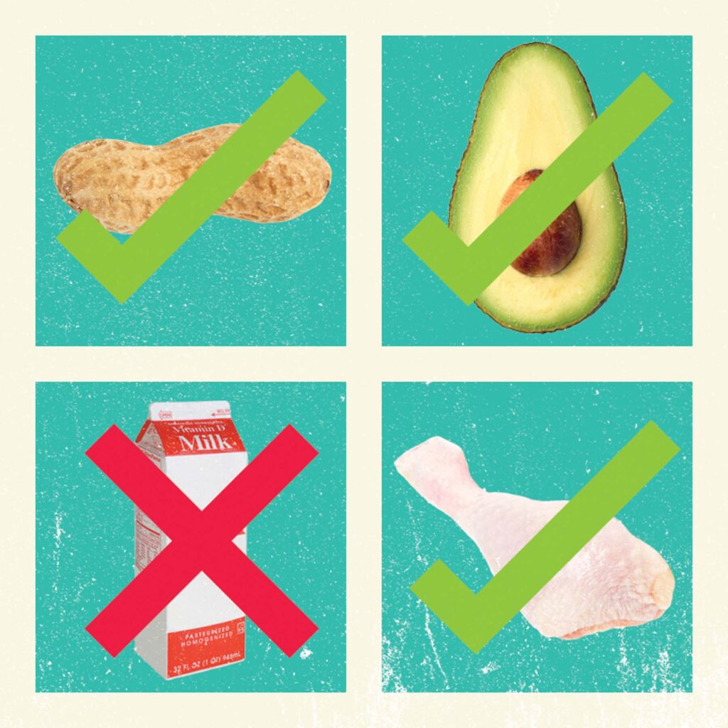
The device works similarly to other detectors commonly found in homes, such as those for carbon monoxide, but it can identify multiple gases instead of just one. That’s thanks to an array of 16 tiny gas sensors that act like finely tuned digital taste buds. The brain behind the nose is a machine learning model trained to recognize how the sensors respond to everything from freeze-dried strawberries to peanuts to raw chicken left out at room temperature for two days.
The e-nose is “far more sensitive and far more objective than any human nose can be,” says study lead Carla Bassil, M.S. ’25, a Ph.D. student in the Department of Electrical Engineering and Computer Sciences. She found that it was able to achieve an overall accuracy of 92.6 percent in classifying 16 different food objects. Beyond spoiled items, the nose could also smell and recognize just 0.05 grams of isolated walnut, a common food allergen.
Bassil will next test the device in environments where other gases are present, such as when sour milk is sitting next to fresh produce in a refrigerator. If it is successful, she says, the e-nose could eventually be integrated into refrigerators themselves. Some already track temperature and inventory, so why not food freshness too.
Cal’s Coral Nursery
 (Brandon Sanchez)
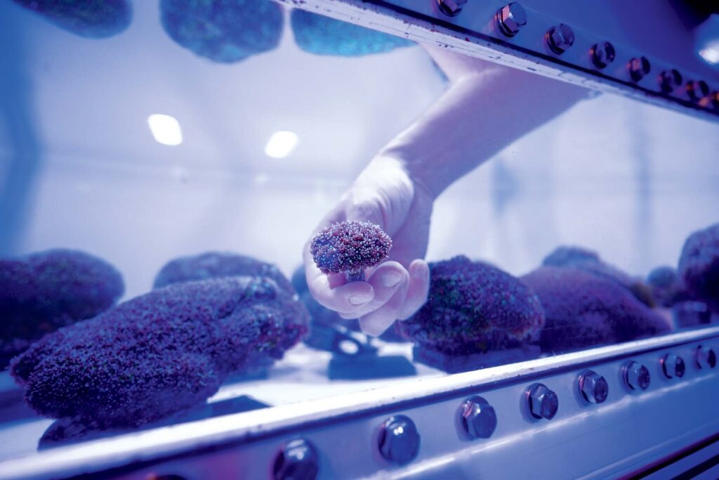
In the basement of Koshland Hall, luminous aquariums house green coral and anemones that resemble translucent palm trees. Here, in Cal’s one-of-a-kind coral nursery, these cnidarians are thriving. Even if they are a little jetlagged.
In their native reefs, most coral only spawn once a year, en masse around a full moon, limiting scientists to a narrow window for collection. By carefully altering his lab’s lighting and temperature, assistant professor Phillip Cleves, Ph.D. ’15, has successfully designed six tanks of coral to spawn sequentially, giving him six opportunities a year to experiment on coral eggs. Their genetics may reveal the answer to how coral evolved with algae, a symbiotic relationship dating back hundreds of millions of years.
His theory: Microscopic algae parasitized coral, learning how to live within the cells’ lysosomes without being eaten and instead releasing the products of photosynthesis to nourish their host, thus creating symbiosomes. When his team broke down some of the 200 symbiosomal proteins in the Aiptasia anemone, a cousin to coral, they found that many of them help keep the algae alive. “There are several vesicle trafficking proteins that we think are actually how the animal and the algae communicate, because they are trafficking cargo on and off the symbiosome, bringing stuff to the algae,” Cleves said. They even mutated one, a bicarbonate transporter in the coral, with CRISPR, possibly cutting off the carbon dioxide the algae needs for photosynthesis.
“Our overall research goal,” he told Berkeley News, “is to understand symbiosis and also to understand the genetic mechanisms for why corals bleach,” the process when stressed coral expel the algae that give them both color and the food needed to survive.
CRISPRing Cancer
 (iStock)
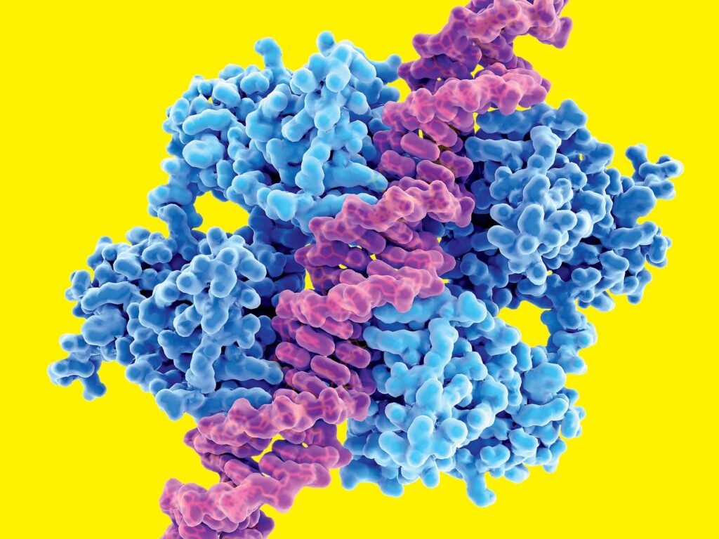
Fourteen years after Berkeley biochemist and Nobel laureate Jennifer Doudna co-created CRISPR-Cas9, researchers in her lab at Berkeley’s Innovative Genomics Institute (IGI) are using the gene-editing technology to target “undruggable” cancers.
Their new technique comes from the long-held understanding that roughly half of all cancers, including 70 to 90 percent of the most stubborn ones (like pancreatic or ovarian), harbor the same faulty protein: p53, a tumor suppressor nicknamed the “guardian of the genome.” When p53 proteins are too mutated to do their job, the cells they’re meant to protect are left vulnerable, allowing tumors to grow.
Scientists have been working on reviving or replacing mutated p53 proteins since the ’90s. But no treatment has ever been approved for clinical use. Part of the problem is that p53 lacks the “pockets” that drug molecules normally latch onto.
That’s where CRISPR comes in. Instead of trying to fix cells with a mutated p53, scientists in Doudna’s lab are using a specialized form of CRISPR to kill them so healthy cells can take their place. “When people treat cancer with chemotherapy or radiotherapy, that’s essentially killing all the dividing cells in the body, including healthy cells,” said Jingkun Zeng, the study lead and a postdoctoral researcher in Doudna’s lab, in an IGI press release. “With this technology, it’s much, much more precise.”
Zeng and team have already succeeded in using CRISPR-Cas12a2 to sort through cultures of mammalian cells, find the mutated few, and shred their genetic material. The technique also promises to extend beyond p53. In Doudna’s words, “Not only can this approach target the ‘undruggable’ cancers that we know, we can also easily and quickly adapt this to new mutations.”
Arresting Cardiac Arrest
 (Iron Studios)
The medical community calls sudden cardiac arrest a “silent killer” for a reason: When the heart suddenly stops beating, few survive the following minutes. Every year, it kills more than 300,000 people in the U.S. alone—very often with no prior warnings or signs of disease.
The solution is simple, says Ziad Obermeyer, a physician and an associate professor at Berkeley’s School of Public Health: Get a defibrillator implanted to regulate your heart. “The problem,” he says, “is that doctors can’t figure out who needs one before it’s too late.”
So Obermeyer began looking to AI for help. Through two organizations he cofounded, Dandelion Health and Nightingale Open Science, he collected six years of EKG scans from hospitals across Sweden. With that data, he trained an AI model to analyze the waveform patterns of both healthy and at-risk hearts. One decade and 440,000 EKGs later, it has identified a biomarker that precedes cardiac arrest.
The model consistently outperforms current detection methods. Where a standard clinical test isolates a patient group with a 4.6 percent annual rate of sudden cardiac death, Obermeyer’s tool can identify groups with a 7 percent rate. Morbid as that may sound, those few percentage points could mean thousands of saved lives.
Currently, Obermeyer is deploying the algorithm in hospitals in Sweden, Taiwan, and the U.S., where it will flag high-risk EKG scans. He also built a website where individuals can sign up for the chance to have their own EKGs analyzed.
With AI, Obermeyer says, “there is going to be a new way of doing science.”
Who Runs the Negotiation? (Girls)
 (iStock)
Earlier research has suggested that women are worse negotiators than men because they’re too accommodating. But “scholars have been so focused on who wins the negotiation that we missed who wins the relationship,” Berkeley behavioral scientist Laura Kray says.
As part of a revealing new paper, Kray, Haas assistant professor Solène Delecourt, and lead author and former graduate student Charlotte Townsend, M.S. ’21, Ph.D. ’24, conducted five different studies with more than 2,000 people and found that, in negotiations, women achieved economic outcomes “on par” with men while simultaneously outscoring them in likeability, trustworthiness, and fairness.
One of the studies drew from anonymous online negotiations where participants haggled over fictitious resources via text chat. Even when the researchers assigned random gender labels to the chats and showed them to hundreds of reviewers, women were ranked highest on all measures of subjective value. They were better liked because of their behavior—not because of gender-based stereotypes. As the paper concludes, people simply prefer negotiating with women.
Over time, that turns into more money. After running a randomized simulation on their data, the researchers estimated that women could see 45 percent more negotiation opportunities over a career. If every negotiation opportunity earned a bonus, for example, that would mean extra dollars in their pockets.
“In personal finance, the value of compounding is extremely powerful in the long term,” Delecourt told UC Berkeley Haas. “The same is true in negotiation: If people enjoy the process of negotiating with you, and want to negotiate with you in the future, that is like a high interest rate. It is likely to result in long-term gains.”
Botanical Pompeii
 (Jaemin Lee)
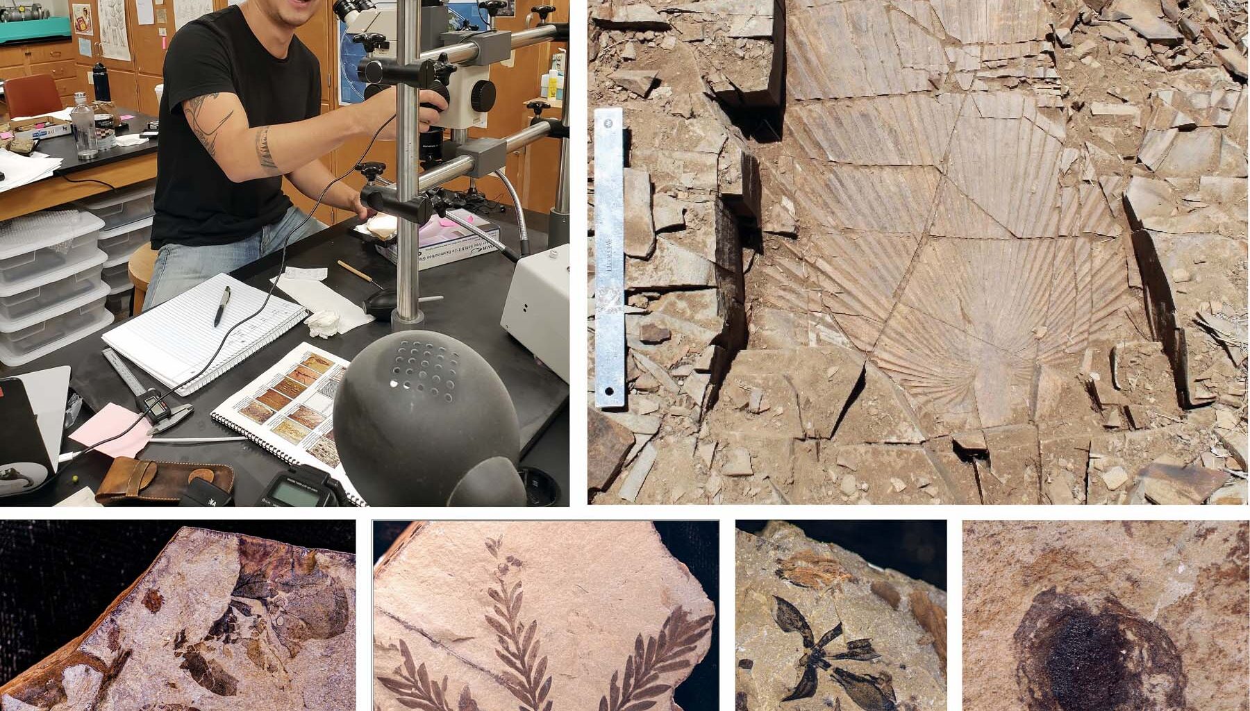
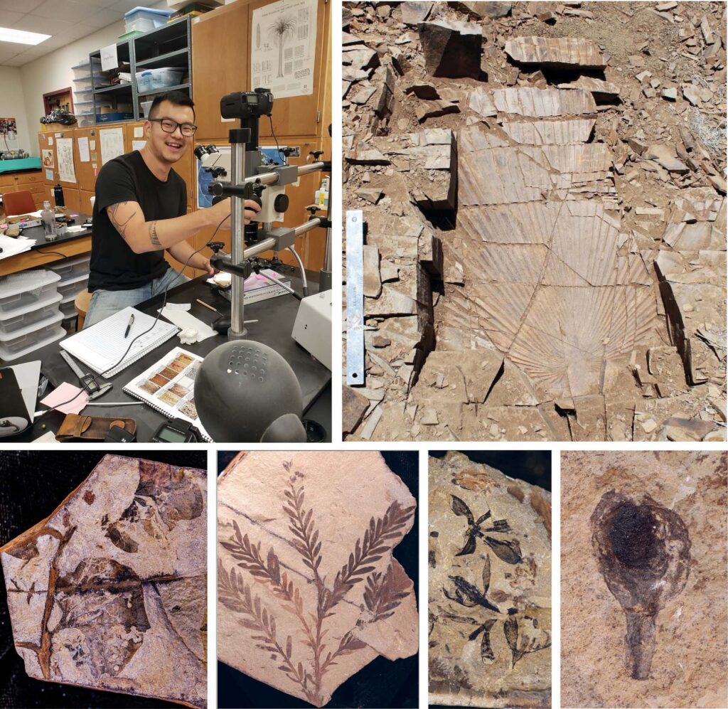
When Mount Vesuvius erupted in 79 AD, volcanic ash buried Pompeii, preserving the city in remarkable detail. Now Berkeley paleobotanists have unearthed a different kind of Pompeii—this one from the time of the dinosaurs.
The 75-million-year-old site, located in New Mexico and known as Dori’s Tuff, was once a thriving tropical forest. Then a nearby volcano erupted, blanketing the inland forest in ashfall and capturing the shapes of plants in solid rock, creating what UC Museum of Paleontology curator Cindy Looy calls “a snapshot in time.”
After excavating thousands of fossilized leaves, flowers, fruits, and seeds, Looy, doctoral student Jaemin Lee, and the Perot Museum of Nature and Science’s Dori Contreras, Ph.D. ’18—for whom the site is named—reconstructed a forest dominated by flowering plants growing alongside older lineages of ferns and redwoods. This upends the common narrative that angiosperms did not start flourishing until after dinosaurs went extinct. What’s more, they found that the seeds and fruits, called diaspores, were unexpectedly large, on average the size of a plump blueberry.
That might not seem like much today, but it’s a hundredfold difference in volume from the poppy seed–sized diaspores of other Cretaceous sites. Rather than dispersing small seeds unassisted, the findings imply that flowering plants were already interacting with early mammals and even dinosaurs in some areas well before the end-Cretaceous extinction.
“This fossil flora suggests that these animals were already moving over to eating bigger seeds produced by angiosperms 75 million years ago. This is a surprise, because people thought they didn’t exist yet,” Looy said. “And here they are.”