
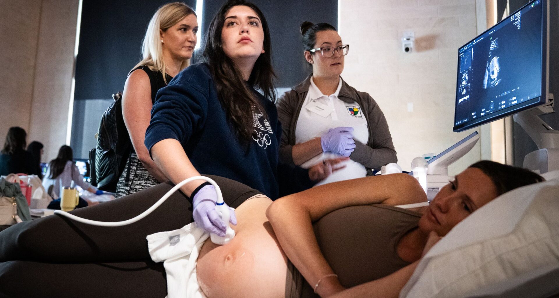
Instructors and nurses gather around a sonogram to watch each other practice a technique during a Tiny Hearts Project training on detecting congenital heart diseases at Dell Medical School.
Sara Diggins/Austin American-Statesman
For many expectant parents, the ultrasound at around 20 weeks of gestation is a celebration: a look at a baby that now has recognizable features, the chance to learn its sex and a checkmark that it appears healthy. Sometimes family members crowd the sonogram room for the occasion.

Parents expect that if anything is wrong with the baby, it will be found; especially if something is wrong with the heart.
Article continues below this ad
Patient Maggie Peterson, a model for the training, looks up at a sonogram scan of her baby during a Tiny Hearts Project training.
Sara Diggins/Austin American-Statesman
Clinicians practice different sonogram techniques during a Tiny Hearts Project training on detecting congenital heart diseases at Dell Medical School.
Sara Diggins/Austin American-Statesman
The display on the sonogram indicates monitoring the mitral valve during a Tiny Hearts Project training on detecting congenital heart diseases at Dell Medical School in Austin, July 11, 2026.
Sara Diggins/Austin American-Statesman
The truth, though: Most congenital heart problems are not found on an ultrasound, even though they can be detected during the exam, said Dr. Celeste Sheppard, a maternal-fetal medicine specialist at Dell Children’s Medical Center. Many sonographers and even obstetricians have not been trained on the scanning and interpreting needed to diagnose a congenital heart problem, Sheppard said.
She started the Tiny Hearts Project nonprofit in 2016 to train more doctors and sonographers on how to better perform the ultrasound anatomy exam and what to look for.
This month, Tiny Hearts Project held a two-day course for obstetricians and sonographers that presented both its Five Essential Views training and other updated ultrasound techniques. It has three more one-day trainings in Austin this year and a training in Weslaco in September.
Article continues below this ad
Yoselin Garcia, center practices a sonogram technique on Maggie Peterson as instructors Lindsay Wood, left, and Jessica Glandt look on during a Tiny Hearts Project training on detecting congenital heart diseases at Dell Medical School in Austin, July 11, 2026. The Tiny Hearts Project aims to train clinicians to be able to detect congenital heart defects early through better training, promoting better outcomes for mothers and babies.
Sara Diggins/Austin American-Statesman
Saving babies through training
Heart problems are some of the most common congenital conditions, affecting 1 in 100 babies, Sheppard said. Many are not life-threatening, but about 3 in 1,000 are, she said. Ultrasounds are only catching about one-third of those, she said. “We need to do better,” Sheppard said.
Congenital heart conditions can range from a tiny hole in the heart to ventricles that are not fully formed, or blocked or abnormal circulation. These problems can prevent adequate oxygen from being delivered throughout the body. Babies can have lips, fingers and toes that look blue or their whole body can look blue because of the lack of oxygen.
Article continues below this ad
When a serious heart condition is diagnosed in the womb, parents and the medical team can arrange for the babies to be born in a hospital with the highest level of cardiac care so the physicians who will be treating the baby can plan for care after delivery, including possible immediate surgery.
“Babies who don’t get detected often are OK the first few hours or days,” Sheppard said. They might even go home, but then, as their circulation gets used to being in the world, they begin to get sicker and sicker.
The missed diagnosis can lead to a baby who is more fragile, needs more intensive and longer care, and might or might not survive, she said. Catching the problem in the womb helps doctors know how to stabilize the baby at birth, preventing the baby from becoming sick from a missed diagnosis.
Dr. Celeste Sheppard, founder of the Tiny Hearts Project, speaks with training providers during a training on detecting congenital heart diseases. Her goal is to have more sonographers and obstetricians trained on finding the five essential views of the heart to find defects.
Sara Diggins/Austin American-Statesman
Learning by doing
Two classrooms at Dell Medical School were set up with ultrasound machines and exam room beds. Pregnant models rested on the beds as participants rolled the ultrasound probe over the models’ bellies to find the five different scans of the heart needed. An ultrasound expert stood beside each trainee, giving tips on which probe to use, when to zoom in, which images to save and when to record the motion of the blood flow through the heart.
Article continues below this ad
They worked with the mother and the baby because, unlike doing an ultrasound on a grown adult’s heart, the baby often doesn’t cooperate by staying still and resting on its back. The trainees asked the model to turn on her side or push on one side of her belly to get the baby to turn.
A sonogram indicates blood flow with colors during a Tiny Hearts Project training on detecting congenital heart diseases at Dell Medical School in Austin, July 11, 2026. The Tiny Hearts Project aims to train clinicians to be able to detect congenital heart defects early through better training, promoting better outcomes for mothers and babies.
Sara Diggins/Austin American-Statesman
Casey Davis, a sonographer in Lufkin, recently switched from doing vascular ultrasounds on adults in a hospital to working at an obstetrics and gynecology office. She took the class to get more training in this kind of ultrasound.
“We want to make sure we’re doing our best and figuring out if there’s something wrong, especially in our area where they would need to be delivered somewhere else,” Davis said. “I want them to get the care they need.”
Article continues below this ad
Davis is exactly the kind of sonographer Tiny Hearts Project is trying to reach, Sheppard said. Tiny Hearts hopes to bring this training to more rural areas. Weslaco is its first expansion to the Rio Grande Valley.
This kind of training can make a big difference to the families whose babies are being scanned.
Whitney Garcia practices a sonogram technique on Devin O’Neill as instructor Dr. Elena Sinkovskaya guides her hand during a Tiny Hearts Project training. Garcia’s son was born with a heart problem that was missed on the original ultrasound. She now works as a sonographer and wants to help more heart defects be caught at the 20-week anatomy scan.
Sara Diggins/Austin American-Statesman
Whitney Garcia, a sonographer who took the training, was one of those parents. Her son, Tobias, now 16 months old, was diagnosed with a coarctation of the aorta on an ultrasound, but it was missed at the initial anatomy scan. She saw a maternal-fetal medicine doctor because her son was conceived by in vitro fertilization. That’s when her son’s heart condition was found.
Article continues below this ad
She and her doctors were able to plan for his birth, including preparing for a possible surgery.
Her son ended up not needing surgery, but the experience made her appreciate the role a well-trained sonographer can have on a baby’s outcome. After Tobias’ birth, she switched from doing general sonography to obstetric sonography.
“I want to learn,” she said. “I don’t want to mess up on anybody’s heart, especially since my son was one of them.”
Article continues below this ad
Instructor Jessica Glandt smiles as she instructs on a sonogram technique during a Tiny Hearts Project training on detecting congenital heart diseases at Dell Medical School. Each ultrasound station had a expert like Glandt to help trainees.
Sara Diggins/Austin American-Statesman