
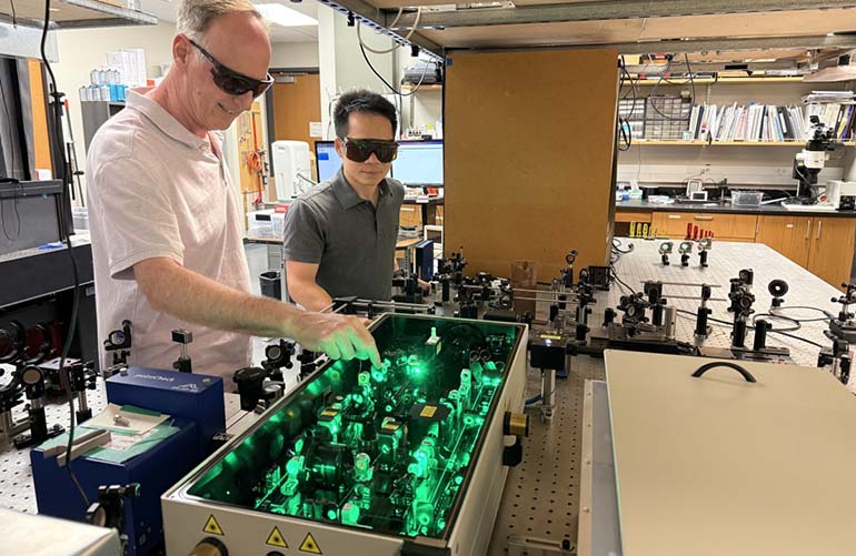
University of Texas at Austin researchers developed a new way to monitor brain blood flow during surgery with a technology called sinusoidal intensity modulation speckle imaging (SIMSI). [Photo courtesy of UT Austin]
University of Texas at Austin researchers have developed a new way to monitor brain blood flow during surgery with standard camera hardware.
The technology is called sinusoidal intensity modulation speckle imaging (SIMSI). It could also be used to improve monitoring during “cardiac surgery, help assess tissue viability in reconstructive procedures, guide treatment decisions in stroke care, and support research into conditions ranging from dementia to traumatic brain injury,” the university said.
“This has been a fundamental challenge in the field for a long time,” UT Austin professor Andrew Dunn said in a news release. “SIMSI gives us a way to get quantitative, physically meaningful numbers from a technique that is already fast and practical enough to use in the clinic.”
SIMSI uses the physics of dynamic light scattering to image blood flow noninvasively across a wide field of view without high-speed cameras. Dunn, who’s also a co-author of the study, leads the lab that published the findings in the Proceedings of the National Academy of Sciences.
Currently available technology requires expensive and specialized high-speed cameras to measure brain blood flow with fast dynamic processes with dynamic light scattering over wide fields.
Register now: The MDO Min-Vasive Medtech webinar series returns Aug. 18-20
Tracking blood flow through the microvasculature during surgery is one of the most critical parts of a procedure, the university said. Interruption in flow could lead to permanent damage, but access to monitoring technology is limited due to high-cost hardware.
SIMSI builds on an established technology called laser speckle contrast imaging (LSCI). LSCI uses laser light to illuminate tissue and the movement of red blood cells causes the laser speckle pattern to blur in proportion to how fast the cells are moving. Researchers then analyze the blur to noninvasively map blood flow across an entire field of view.

University of Texas at Austin professor Andrew Dunn [Photo courtesy of UT Austin]
LSCI relatively measures whether blood flow increased or decreased, but doesn’t provide figures for absolute blood flow. The researchers designed a camera-based system that noninvasively produces quantitative maps of rapid blood flow dynamics across a wide field of view, the university said.
The researchers added precise modulation to the laser illumination by varying light intensity in a sinusoidal pattern at a controlled frequency within each camera exposure, the researchers said. They overcame hardware limitations by encoding information about fast blood flow dynamics into images captured with long camera exposures.
“Blood flow can change over many timescales, but the optical fluctuations that reveal how fast blood is moving are often too fast for standard cameras to sample directly,” said postdoctoral fellow and SIMI development leader Hengfa Lu.
“By intentionally modulating the illumination during the exposure and using a newly derived imaging model, we can recover fast blood flow dynamics that would otherwise be averaged out,” he continued. “That gives researchers a more quantitative way to study how tissue is functioning, from stroke recovery in the lab to, eventually, surgical decision-making.”
The researchers see SIMSI as a broader effort to make fast, quantitative blood flow imaging more accessible. The physics-based imaging model that uses standard cameras could open opportunities in “stroke research, brain injury, surgery and other areas where blood flow is an early signal of tissue health,” UT Austin said.