
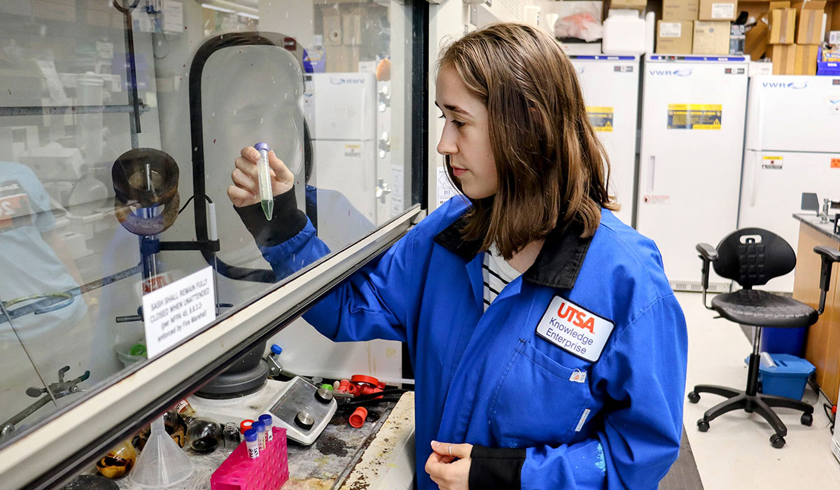
 Syed Muhammad Usama
Surgical removal of a tumor remains the primary defense against most solid cancers. However, for surgeons in the operating room, differentiating between the outer edges of a tumor and healthy tissue is a challenge. In aggressive, structurally erratic cancers, this boundary can be especially hard to find. Leaving behind even a microscopic cluster of residual cancer cells significantly elevates the risk of the disease returning and spreading throughout the body.
Sadly, surgeons are not afforded a perfect map of the tumor and instead, must rely on limited visual cues and touch. But a UT San Antonio College of Sciences researcher is working to provide that map.
Syed Muhammad Usama, PhD, assistant professor of chemical biology in the Department of Chemistry, is developing a novel molecular “smart probe.” The technology is designed to make hidden cancer cells glow in real time during surgery, giving surgeons cleaner margins and patients lower risk of recurrence.
The clinical potential of his work has earned funding from the Max and Minnie Tomerlin Voelcker Fund, including a recent award of $450,000. The award will help accelerate the project from basic organic chemistry into translational models that could reshape the surgical oncology landscape.
Overcoming the blind spots of breast cancer surgery
Usama’s research focuses on triple-negative breast cancer, an exceptionally aggressive subtype that constitutes roughly 15% to 20% of all breast cancer cases. The phrase “triple-negative” refers to the notable absence of the three receptors typically found in breast cancer cells: estrogen, progesterone and human epidermal growth factor receptor 2, or HER2.
These three receptors are commonly the targets that breast cancer therapies and hormone treatments would latch onto. Without them, triple-negative breast cancer is resistant to many first-line treatments.
“Surgical resection is the primary intervention for more than half of all cancer patients every year,” Usama said. “But when you are dealing with a subtype as aggressive as triple-negative breast cancer, relying solely on a surgeon’s sight and touch to clear the tumor margins is a major clinical challenge.”
Fluorescence-guided surgery is an emerging approach that injects patients with fluorophores — targeted fluorescent dyes — prior to an operation. During surgery, specialized near-infrared lasers illuminate these dyes, giving the medical team a glowing blueprint of the tumor and its borders.
Unfortunately, today’s fluorescent dyes also need to bind to the three receptors that triple-negative breast cancer lacks.
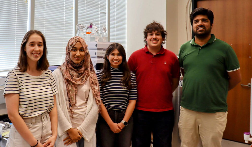 Left to right: honors college students: Lauren Wolff, Rabia Haque, Alyssa Kornegay, College of Sciences student Stefan Nashawati, and Syed Usama.Turning an internal protein into a beacon
Rather than searching for markers on the outside of the cell, Usama’s laboratory is targeting proteins in the cells that help fuel tumor growth from the inside. These proteins are known as cyclin-dependent kinases 4 and 6 (CDK 4/6).
While normal cells keep these proteins relatively controlled, most triple negative breast cancer tumors produce much more of them.
To target these cells, the researchers are pairing an FDA-approved drug called palbociclib (pal), which naturally binds to CDK 4/6, with a deep-tissue-penetrating near-infrared fluorogenic dye called cyclizing heptamethine cyanine (c-Cy7), which Usama developed. Working in tandem, the two compounds form a sophisticated “smart probe” called c-Cy7-pal.
What sets this probe apart is its “smart” activation mechanism. Conventional imaging dyes are “always on,” glowing continuously from the moment they are injected. This creates background noise because the dye pools in healthy tissues, making it hard for surgeons to see the edges of the tumor.
But Usama’s newly engineered probe acts like an intelligent warning light, illuminating cancerous tissue and going dark elsewhere.
“Our c-Cy7-pal probe only activates and illuminates after binding to the specific intracellular proteins overexpressed in the tumor. This gives us dramatically higher target-to-background contrast, making a clear distinction between tumor and healthy tissue,” Usama explained.
This switch-flipping is triggered by changes in the target area’s internal chemistry — specifically how water-repelling or electrically charged the surrounding environment is. While traveling through healthy tissue, the probe naturally twists into a closed, dark state. When the probe penetrates a cancer cell and locks onto the targeted CDK 4/6 protein, the shift in polarity coaxes the molecule to snap open into a charged state, switching on its fluorescence.
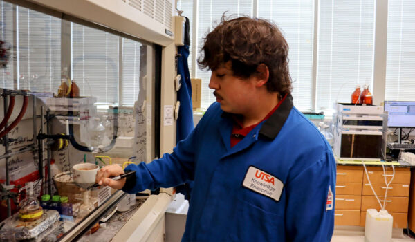 Chemistry research assistant Stefan Nashawati drawing reaction schemes for development of NIR-II fluorophores.
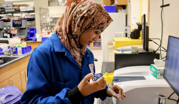 Chemistry major and undergraduate researcher Rabia Haque taking optical properties of near-infrared fluorophores.Training the next generation of STEM leaders
The project is part of UT San Antonio’s broader mission to cultivate an agile STEM workforce in South Texas.
With support from the Voelcker Fund Young Investigator Award, Usama has recruited and trained five PhD students and 12 undergraduate students in chemistry, biochemistry, biology and biomedical engineering. He has redesigned junior-level biochemistry coursework and launched a specialized graduate-level chemical biology course to further expand student access to hands-on laboratory training in molecular design, drug development and bioimaging.
“Our students aren’t just reading about these concepts — they are actively synthesizing the probes, running the biological assays and learning how to communicate complex science. To see their growth has been tremendous; they are the medical and scientific leaders of the future,” Usama said.