

Nikon has unveiled the winners of its 51st annual Nikon Small World Photomicrography Competition. The top 20 winning images showcase the best microscopy and extreme macro photographs of the year.
Zhang You Wins First Place for a Stunning Rice Weevil Portrait
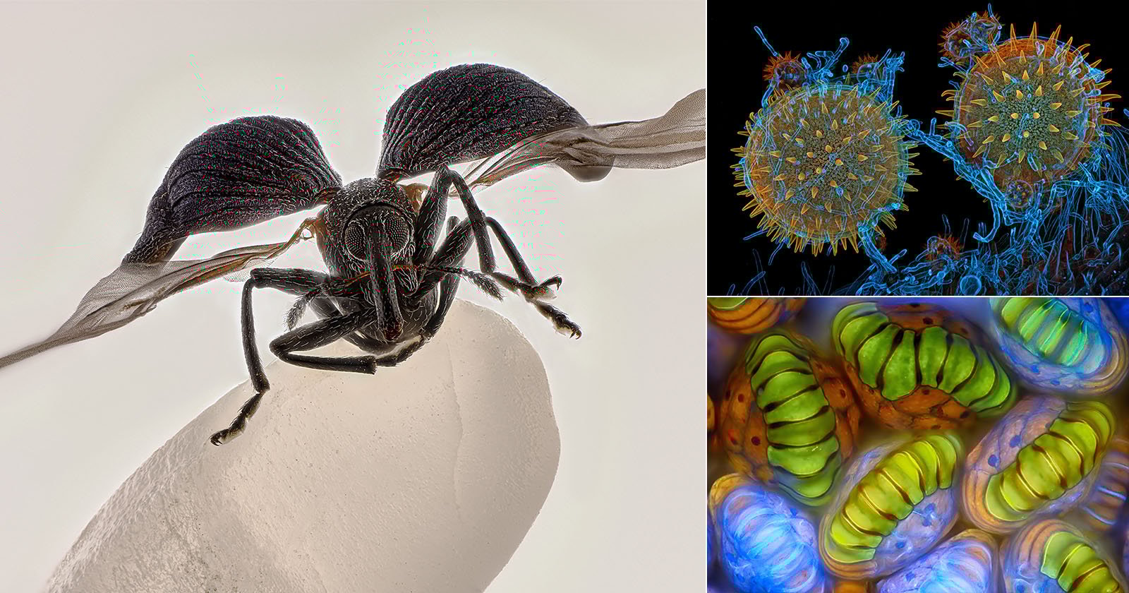
This year’s first-place winner is Chinese photographer Zhang You, whose photo of a rice weevil mounted on a grain of rice shows a common agricultural pest in a striking way, providing a new perspective on an often overlooked insect. You’s photo shows the rice weevil with its wings fully extended, frozen in time.
 1st Place — Rice weevil (Sitophilus oryzae) on a grain of rice by © Zhang You. Image stacking, 5x (objective lens magnification). | Nikon Small World
You is a member of the Entomological Society of China and the Entomological Society of Yunnan Province. Nikon notes that the winning photo is the culmination of You’s many years of dedicated ecological and insect science photography. You is also an entomology teacher.
“It pays to dive deep into entomology: understanding insects’ behaviors and mastering lighting,” the photographer explains. “A standout work blends artistry with scientific rigor, capturing the very essence, energy, and spirit of these creatures.”
The scale of the winning photo is fascinating. While the insect appears to be a fairly typical size, given that it is on a grain of rice, it is clear that the rice weevil is actually tiny. You used a medium-format camera and a 5x microscope objective, and ultimately stacked over 100 photos to achieve the expanded depth of field. It took two weeks of careful shooting and post-processing to create the final image.
“I had observed rice weevils in grains before, but never one with its wings spread,” You says. “This one was naturally preserved on a windowsill, perhaps in a final attempt to escape. Its tiny size makes manually preparing spread-wing specimens extremely difficult, so encountering it was both serendipitous and inspiring.”
In addition to his victory, Zhang You also took 15th place in the competition, and this image is seen further down.
“Zhang You’s work demonstrates the remarkable power of microscopy to reveal new perspectives on the world around us,” says Eric Flem, Senior Manager, Communications and CRM at Nikon Instruments.
“What makes this year even more extraordinary is that it was his very first time entering the competition, and he not only captured first place, but also placed another image in the top 20. His achievement highlights the spirit of Nikon Small World: inspiring wonder, making scientific understanding accessible to all, and celebrating the artistry of the microscopic realm,” Flem continues.
Second and Third-Place Winners 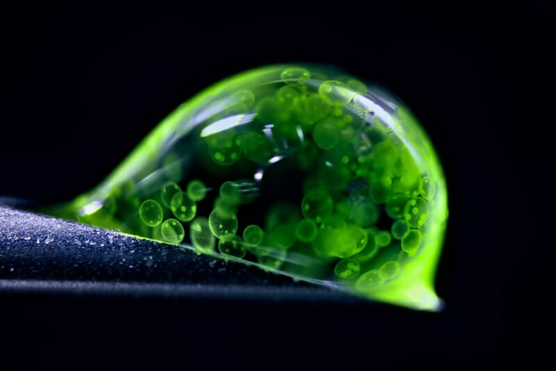 2nd Place — Colonial algae (Volvox) spheres in a drop of water by © Dr. Jan Rosenboom. Reflected light, 5x (objective lens magnification). | Nikon Small World
 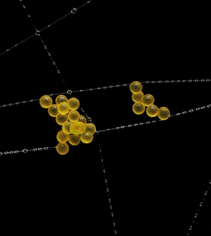 3rd Place — Pollen in a garden spider web by © John-Oliver Dum. Image stacking, 20x (objective lens magnification). | Nikon Small World The Rest of the Top 20
 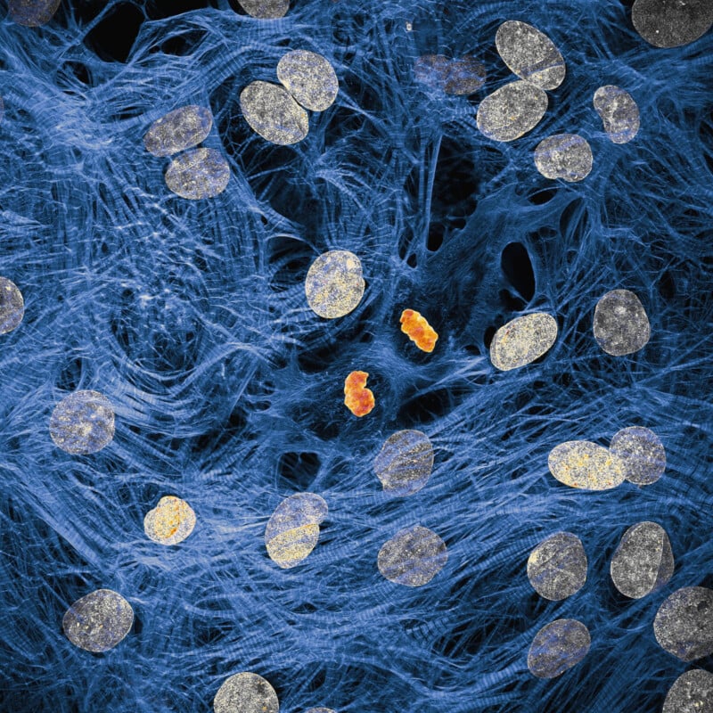 4th Place — Heart muscle cells with chromosomes condensed following cell division by © Dr. James Hayes. Confocal, 100x (objective lens magnification). | Nikon Small World
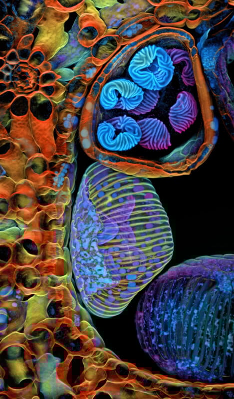 5th Place — Spores (blue/purple structures) of a small tropical fern (Ceratopteris richardii) by © Dr. Igor Siwanowicz. Confocal, 25x (objective lens magnification). | Nikon Small World
 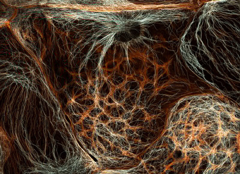 6th Place — Rat liver cells by © Dr. Francisco Lázaro-Diéguez. Confocal, 63x (objective lens magnification). | Nikon Small World
 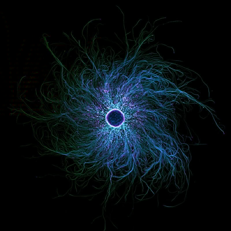 7th Place — iPSC-derived sensory neurons labelled to show tubulin and actin by © Stella Whittaker. Confocal, fluorescence, image stacking, 10x (objective lens magnification). | Nikon Small World
 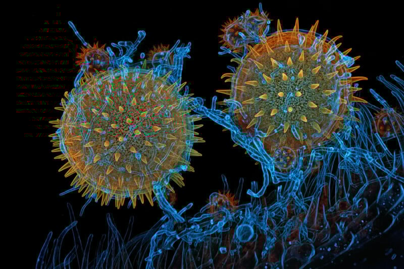 8th Place — Mallow pollen germinating on stigma while being parasitized by a filamentous fungus by © Dr. Igor Siwanowicz. Confocal, 40x (objective lens magnification). | Nikon Small World
 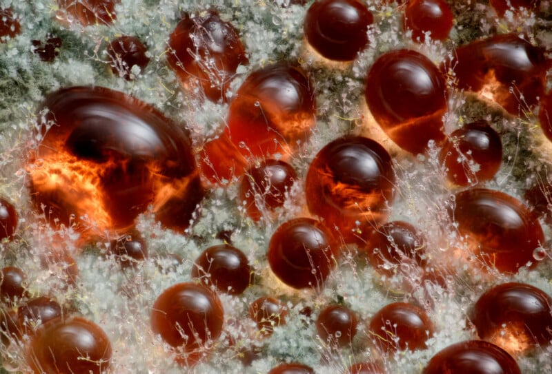 9th Place — A fungus (Talaromyces purpureogenus) known for its red, diffused pigment by © Wim van Egmond. Image stacking, 10x (objective lens magnification). | Nikon Small World
 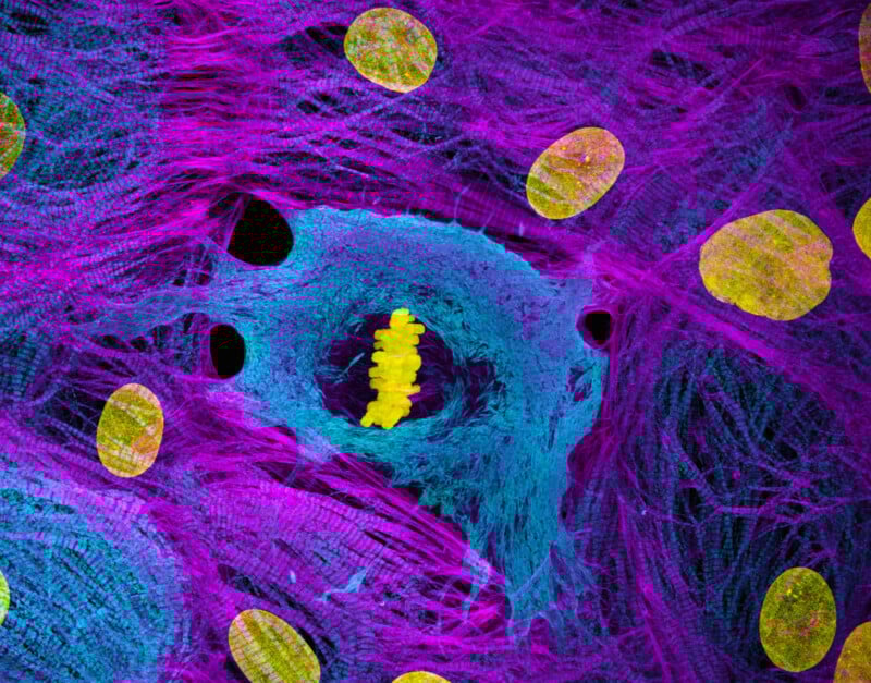 10th place — Heart muscle cells (iPSC-derived) showing condensed chromosomes in metaphase by © Dr. Dylan Burnette & Dr. James Hayes. Structured illumination microscopy (SIM), 60x (objective lens magnification). | Nikon Small World
 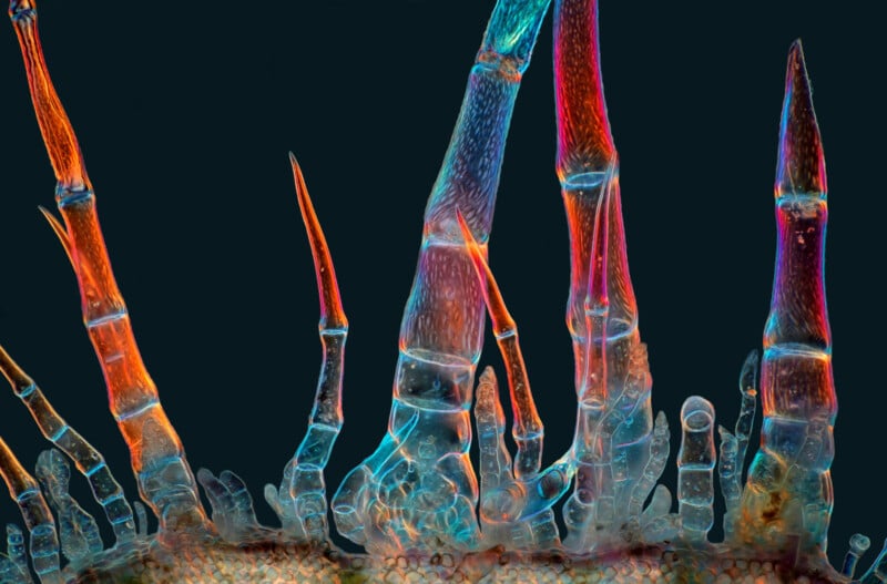 11th Place — Sunflower trichomes (hair-like plant outgrowths) by © Marek Miś. Polarized light, 10x (objective lens magnification). | Nikon Small World
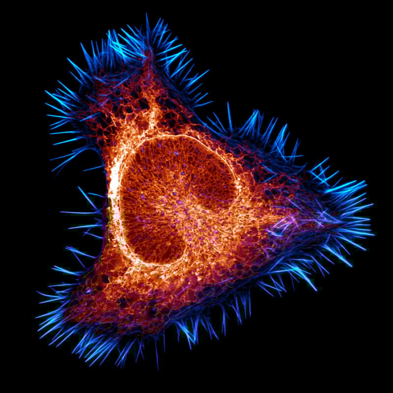 12th Place — The actin cytoskeleton (cyan) and endoplasmic reticulum (red) of a mouse brain cancer cell by © Halil Lindamood & Eric Vitriol. Confocal, deconvolution, 100x (objective lens magnification). | Nikon Small World
  13th Place — Slime mold (Arcyria major) releasing spores by © Henri Koskinen. Image stacking, reflected light, 10x (objective lens magnification). | Nikon Small World
  14th Place — Quartz with biotic goethite filaments by © Manfred Heising. Image stacking, 5x (objective lens magnification). | Nikon Small World
 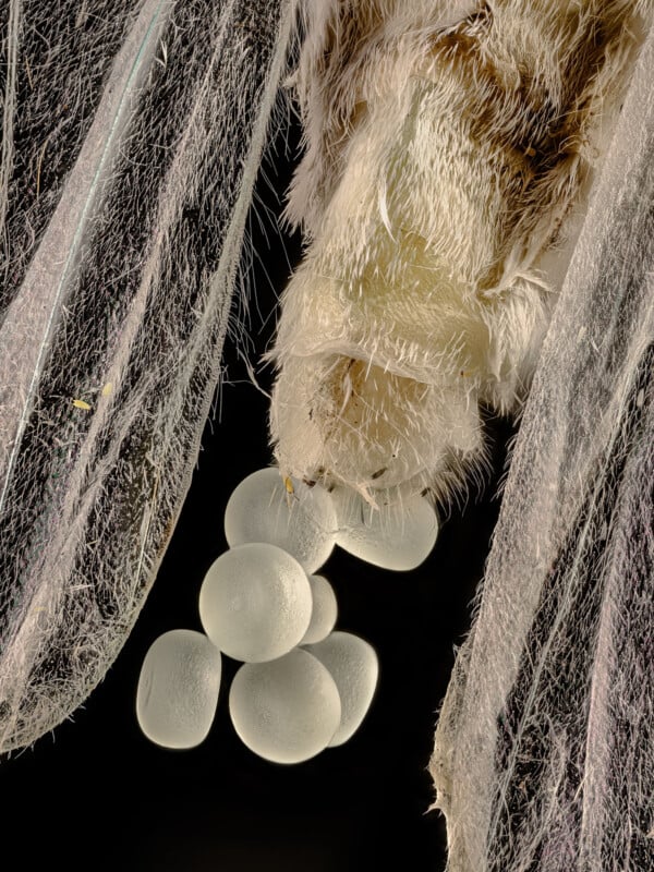 15th Place — Geometer moth (Geometridae) laying eggs by © Zhang You. Image stacking, 5x (objective lens magnification). | Nikon Small World
 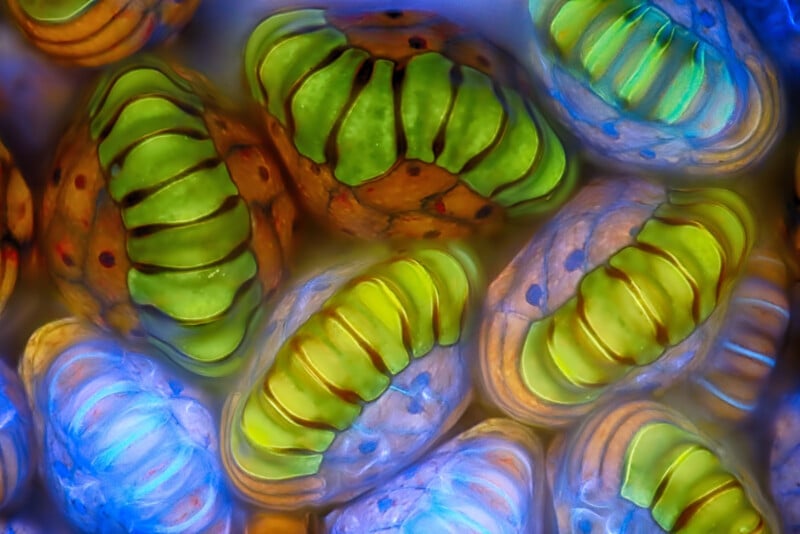 16th Place — Water fleas (Daphnia) and algae by © Rogelio Moreno. Fluorescence, image stacking, 40x (objective lens magnification). | Nikon Small World
 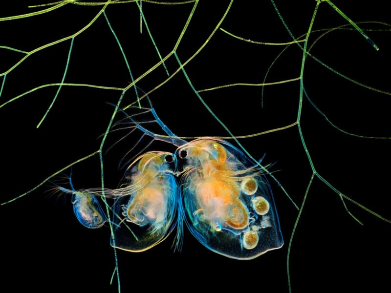 17th Place — Water fleas (Daphnia) and algae by © Hong Guo. Image stacking, 5x (objective lens magnification). | Nikon Small World
 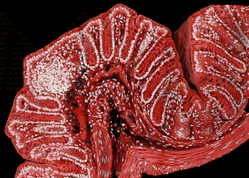 18th Place — Fluorescently marked mouse colon by © Marius Mählen, Koen Oost, Prisca Liberali & Laurent Gelman. Confocal, 20x (objective lens magnification). | Nikon Small World
 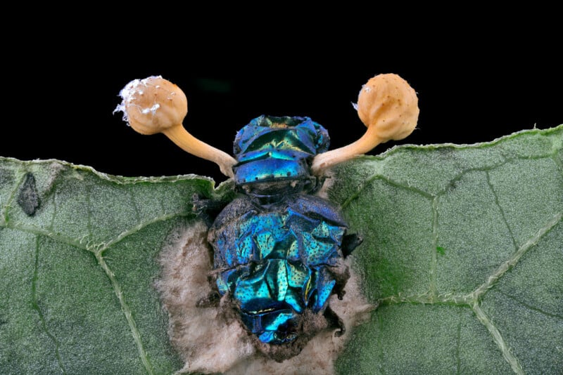 19th Place — Parasitic fungus (Cordycipitaceae) on a fly (Calliphoridae) by © Eduardo Agustin Carrasco. Image stacking, 2x (objective lens magnification). | Nikon Small World
 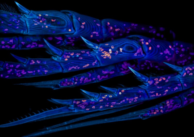 20th Place — Marine copepod by © Zachary Sanchez. Confocal, 60x (objective lens magnification). | Nikon Small World More From the 51st Nikon Small World Photomicrography Competition
Alongside the top 20 images featured above, this year’s judges awarded an additional 51 images for their excellence, including Honorable Mentions and Images of Distinction. These additional winning photos are available on the Nikon Small World website.
Image credits: Photos provided courtesy of Nikon Small World. Photographers are credited in the individual captions.