
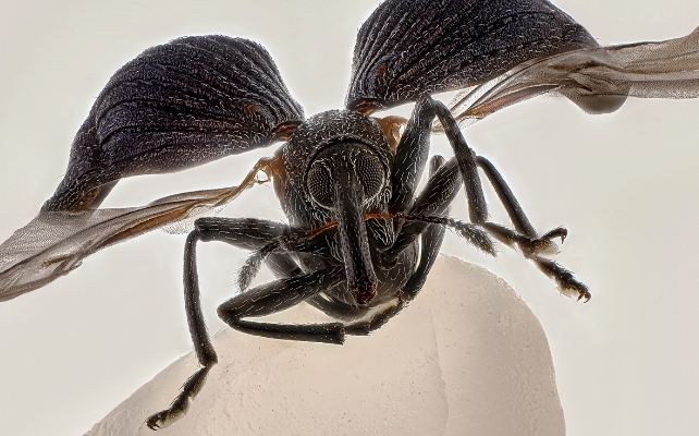
An astoundingly detailed weevil on a single grain of rice takes first place in 2025’s Nikon Small World photomicrography competition.
Entomology enthusiast Zhang You created this intricate image by stacking over 100 carefully lit and cleaned images together.
This technique allows the photographer to maintain a sharp focus across the depth of a subject, whereas a single photo will only be focused across a small range of depth.
The whole process took two weeks.
Related: Award-Winning Image Reveals a Hidden Culprit Behind Alzheimer’s
“It pays to dive deep into entomology: understanding insects’ behaviors and mastering lighting,” says You.
Insects play crucial roles in our living biosphere, from pollination to cleaning up organic waste.
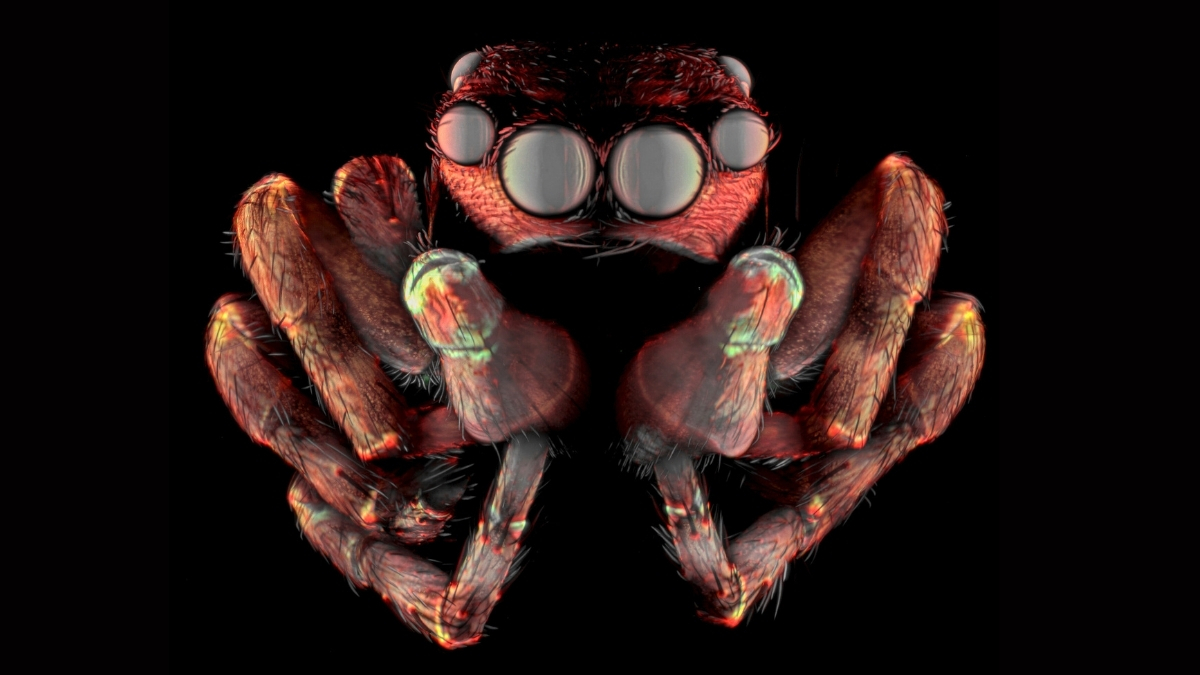
 A cropped version of the winning image reveals some of its incredible details. (Zhang You)
While some insects, like You’s weevil, are considered pests, many species are now declining.
Such powerful imaging can encourage us all to see the incredible complexity of their world and strive to understand them better.
Now in its 51st year, entries to Nikon’s highly specialized photography competition continue to awe us with the intricacies of existence at the microscopic scale.
Similarly precise and time-consuming efforts as You’s likely went into all 1,925 photo entries from 77 countries.
 Here are a few others that caught our eyes.
Tenth place went to a photo of dividing heart cells. When our cells prepare to divide, they bundle up our genetic molecules (our chromosomes, shown in bright yellow) before cellular machinery pulls them into the two parts of the fracturing cell.
We can see an example of this bundling in the image below, where the chromosomes look like a stack of bricks in the middle of these adult heart cells, which have been grown from other adult cells.
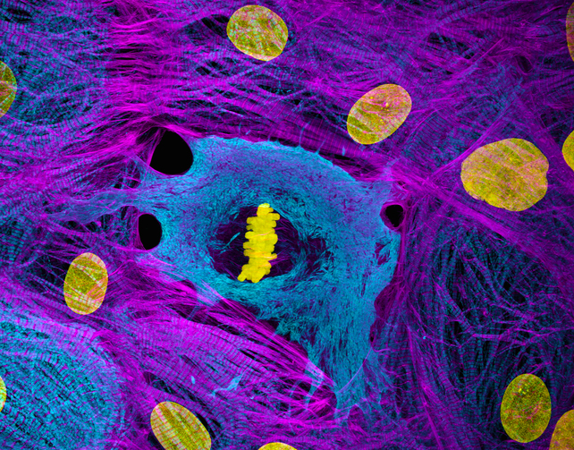 Heart muscle cells derived from induced pluripotent stem cells ( iPSCs) showing condensed chromosomes (bright yellow) in metaphase. Structured illumination microscopy, 60X. (Dylan Burnette & James Hayes/Vanderbilt University School of Medicine)
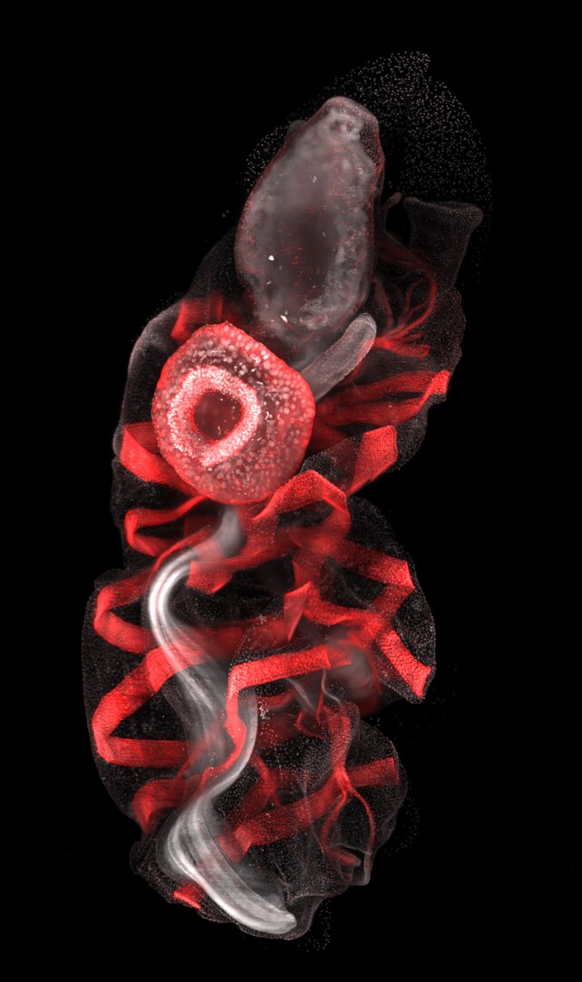
Disembodied vasculature in the shape of a mammalian hand reaches out from the embryo of a developing mouse, in this eerie image, which earned a distinction.
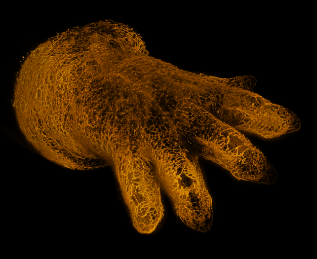 Blood vessels in the limb of an embryonic mouse. Light sheet microscopy, 1X. (Michael Weber/Berlin Institute of Health at Charité)
A disconcerting trip into the strange alternate dimension of soy sauce fused with alum, a combination of hydrated salts and metals often used as a preservative, has us feeling dizzy.
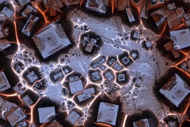 Crystallized soy sauce fusion with alum. Polarized light microscopy, 10X. (Mishal Abdulaziz Alryhan/FIAP)
This magnificent blob is an oozoid, the solitary asexual phase of a sea squirt (Thalia democratica).
When it is ready to reproduce, it will ooze out a long chain of clones, which develop into females.
These females release eggs that become new oozoids, and then turn into males that release sperm to fertilize other neighboring female chains.
 Oozoid of a sea squirt (Thalia democratica). Light sheet microscopy, 10X. (Mette Handberg-Thorsager, Alexandre Alié & Lisa Maria Ulbrich/Georg-August-University Göttingen)
Polarized light brings out unexpected color combinations and structures in this image of recrystallized phenyl imidazol, a substance often used to help create other chemicals.
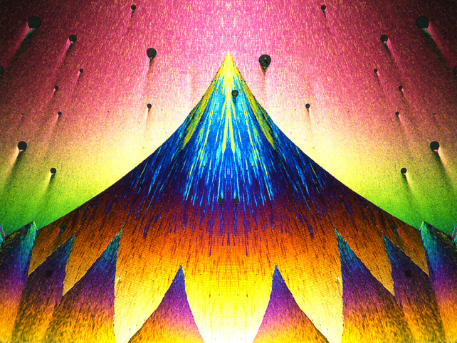 Recrystallization of phenyl imidazol. Polarized light microscopy, 10X. (Karl Deckart)
A swirling galaxy of fluorescently labeled cellular structural proteins, actin and tubulin, outline the intricate branches of a sensory neuron. It’s other-worldly.
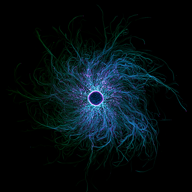 iPSC-derived sensory neurons labelled to show tubulin and actin. Confocal microscopy, 10X. (Stella Whittaker/National Institutes of Health)
These delightfully odd pink latticed hats are the delicate little eggs of a butterfly, Artopoetes pryeri.
It’s a small white and black butterfly found in Asia, and the males have a dusting of blue to purple iridescence.
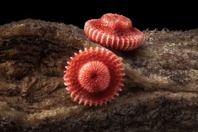 Butterfly (Artopoetes pryeri) eggs. Image stacking, 10X. (Ye Fei Zhang)
You can view many other entrants and winners here.