
The story of life’s beginnings gets stranger when you look closely at viruses. These tiny entities seem to sit at the edge of biology. They carry genetic material, but they cannot make proteins on their own. That single limitation keeps them from acting like independent life.
Still, viruses have likely been around since the first cells appeared. That long history has kept one question alive for decades. Where did viruses come from, and how did they shape the living world?
A new discovery from researchers in Japan adds a fresh clue. The team reports a newly identified giant DNA virus that infects amoebae. They named it ushikuvirus, after Lake Ushiku in Japan’s Ibaraki Prefecture, where they isolated it.
The finding matters because this virus behaves in ways that connect two different viral strategies. It also adds weight to a provocative idea about evolution. That hypothesis suggests viruses may have helped create one of the defining parts of complex cells, the nucleus.
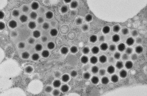
Morphological features of ushikuvirus particles and CPE of infected cells. (CREDIT: Journal of Virology) A Long Debate Over Where the Nucleus Came From
You rely on eukaryotic cells every day. They make up animals, plants, and fungi. These cells have a nucleus wrapped in a membrane. The nucleus stores DNA and helps organize the cell’s work.
Scientists have debated how the nucleus first appeared. Professor Masaharu Takemura from the Graduate School of Science at Tokyo University of Science has pursued one particular possibility for years. In 2001, he and Dr. Philip Bell of Macquarie University independently proposed what is known as the cell nuclear virus origin theory. Dr. Bell also coined the term viral eukaryogenesis.
This hypothesis argues that the nucleus may trace back to a large DNA virus. In this scenario, a virus infected an archaeal ancestor and did not kill it. Instead, it set up a lasting presence in the cell’s interior. Over time, the virus acquired key genes from its host. That long co-existence could have produced the nucleus seen in eukaryotic cells today.
The idea sounds extreme, but giant viruses keep giving it new places to stand. Giant DNA viruses were discovered in 2003, and they surprised scientists with their size and complexity. When these viruses infect cells, they often build “virus factories.” These are specialized structures inside the host where viral DNA replication happens.
Some of those factories sit inside a membrane. That detail catches your attention for a reason. A membrane-bound factory can resemble a nucleus in function and form. It creates a controlled space for copying genetic material.
Morphological features of ushikuvirus particles in infected cell cytoplasm at 5 hpi revealed by c-TEM analysis. (CREDIT: Journal of Virology) A Giant Virus Found in Freshwater
The new study describes a giant DNA virus that infects vermamoeba, a type of amoeba. Vermamoeba are single-celled organisms found in the environment. Ushikuvirus joins a growing list of giant viruses known to infect amoebae.
The research team included Master’s students Jiwan Bae and Narumi Hantori from Tokyo University of Science. It also included Dr. Raymond Burton-Smith and Professor Kazuyoshi Murata from Japan’s National Institute of Natural Sciences.
Takemura framed giant viruses as an open frontier. “Giant viruses can be said to be a treasure trove whose world has yet to be fully understood,” he said. “One of the future possibilities of this research is to provide humanity with a new view that connects the world of living organisms with the world of viruses.”
That sense of discovery comes with a practical problem. Giant viruses appear widely in the environment, but isolating them remains difficult. They are diverse, and researchers do not yet have an easy path to find them all. That makes each new isolate valuable, especially when it behaves differently than its closest relatives.
A Capsid With New Details
Ushikuvirus looks similar, at first glance, to viruses related to the family Mamonoviridae. It also resembles Medusavirus, a genus known for an icosahedral shape and many short spikes on the capsid surface. That outer shell, the capsid, protects the viral genetic material.
But this new virus does not match the mold completely.
A joint research team discovered and characterized ushikuvirus, a new giant DNA virus that infects vermamoeba. This image (a) shows the 3D reconstruction of the virus, highlighting its spiked capsid (d). (CREDIT: Professor Kazuyoshi Murata from the National Institute of Natural Sciences (NINS))
The researchers report that ushikuvirus shows distinct features on its capsid surface. It has multiple spike structures with unique caps. Some spikes include filamentous extensions. The team did not see those features in medusaviruses.
The virus also produces a striking effect on its host. It triggers a cytopathic effect that causes vermamoeba cells to become unusually large. That swollen appearance offers a visible clue that the virus is reshaping its host’s biology.
These differences matter because they help scientists compare viral lineages. Over time, those comparisons can reveal how giant viruses diversified. They can also show how host species may have pushed viruses toward different solutions.
A Different Way to Use the Nucleus
Ushikuvirus also stands out in how it replicates.
Some related giant viruses replicate within the host nucleus while the nuclear membrane stays intact. The source material describes this pattern for medusaviruses and clandestinovirus. Ushikuvirus takes a different approach. It disrupts the nuclear membrane while producing viral particles.
That behavior places it in an interesting middle ground. The researchers suggest it may link viruses that rely on an intact nucleus as a factory with giant viruses, such as pandoravirus, that disrupt the nuclear membrane during replication.
CPE of the host cell size by ushikuvirus infection. (CREDIT: Journal of Virology)
You can picture this as a spectrum of strategies. On one end, a virus uses the nucleus without breaking it. On the other, a virus breaks down the barrier to build what it needs. Ushikuvirus appears to sit between those extremes.
The team suggests these differences may reflect adaptations to hosts. Even closely related viruses can face different pressures depending on which amoeba species they infect.
Takemura expects this discovery to sharpen scientific debate. “The discovery of a new Mamonoviridae-related virus, ‘ushikuvirus,’ which has a different host, is expected to increase knowledge and stimulate discussion regarding the evolution and phylogeny of the Mamonoviridae family,” he said. He added that it could help scientists “get closer to the mysteries of the evolution of eukaryotic organisms and the mysteries of giant viruses.”
Practical Implications of the Research
This discovery gives researchers a new comparison point for how giant viruses interact with cell structures, especially the nucleus. By studying a virus that disrupts the nuclear membrane, scientists can better map the range of strategies used by related viruses. That can deepen discussions about viral evolution and how complex cells may have formed.
The findings also support continued testing of the nuclear virus origin hypothesis. Giant viruses that build factory-like structures inside host cells offer real biological examples of compartmentalized DNA replication. Each newly described virus adds detail that can clarify which features are shared across lineages and which evolved later.
The work may also help inform health research tied to amoebae. The source material notes that certain Acanthamoeba species can cause diseases such as amoebic encephalitis. While ushikuvirus infects vermamoeba, learning how giant viruses infect and destroy amoebae could eventually guide new ways to prevent or treat amoeba-related infections. The benefit would come from understanding the weak points in amoebae biology that viruses exploit.
Related Stories