
Both patients presented with progressive, treatment-refractory autoimmune neuropathy associated with either IgM-kappa paraproteins or MAG-Abs and were at risk of losing ambulatory function.
Patient 1
A 65-year-old woman with a five-year history of IgM-kappa paraproteinemic neuropathy presented with progressive distal sensory loss in the arms and legs, leg and lower back pain, and gait disturbance. Despite previous treatments, including high-dose steroids, IVIG and plasma exchange, her clinical status progressively deteriorated (Supplementary Table 1) and she presented with a reduced maximal walking distance (baseline: 300 meters). She typically necessitated bilateral support walking outdoors, exhibited significant difficulty climbing stairs, and was unable to run or tolerate prolonged standing. As nerve conduction studies demonstrated a sensory-motor neuropathy with marked demyelinating features, while sonography showed pronounced nerve swellings, she was diagnosed with CIPNM and IgM-kappa paraproteins.
Teclistamab induced a rapid clinical response, marked by increased walking distance, enhanced grip strength, and improved neuropathy assessment scores (INCAT, I-RODS) along with significant recovery in nerve conduction velocity, distal motor latency, and compound muscle action potential amplitudes, as well as resolution of nerve swelling. Patient-reported outcomes, including quality of life, pain, and fatigue also improved (Fig. 1, SFig. 1, Supplementary Table 2). In good agreement with the clinical and neurophysiological improvement, serum neurofilament light chain (Nfl) levels also continuously decreased upon treatment with teclistamab, indicating an attenuation of neuronal injury (SFig. 1C). Peripheral blood flow cytometry analysis revealed a transient consumption of T-cells followed by a depletion of B-cells with B-cell repopulation starting approximately four months after teclistamab treatment (Fig. 2A). Repopulating B-cells were predominantly characterized by a naïve phenotype at month 6 and 9 whereas plasmablasts and plasma cells as well as more differentiated B-cells were barely detectable in peripheral blood and did not change substantially (Fig. 2B, SFig. 2). These findings are in line with previous reports on BiTE and CAR T-cell treatment in autoimmune diseases targeting CD1915,16. Concurrently, soluble BCMA (sBCMA), a short half-life biomarker of late B-cells, plasmablasts, and plasma cells17, also decreased rapidly and reoccurred before re-expansion of circulating B-cells post-treatment (Fig. 2C). Interestingly, M-Protein completely disappeared and was undetectable until last follow-up at 9 months (day 287), possibly indicating a polyclonal rather than monoclonal re-expansion of plasmablasts and plasma cells following teclistamab treatment. In line with this, six months after teclistamab treatment, kappa free light chains became detectable again despite the continued absence of M-protein (Fig. 2C). Levels of IgM, IgG and IgA also decreased, while vaccination titers were largely preserved (Fig. 2D, SFig. 3). Adverse events were mild, including a grade 2 cytokine release syndrome (CRS), neutropenia, and hypogammaglobulinemia, which were well controllable with a single administration of tocilizumab and granulocyte colony-stimulating factor (G-CSF), and two IVIG infusions, respectively (Fig. 2E). The positive clinical and paraclinical response was maintained without signs of disease reoccurrence until the last follow-up (month 9).
Fig. 1: Patient 1: Clinical responses and patient-reported outcome in refractory IgM-paraproteinemic autoimmune neuropathy treated with teclistamab.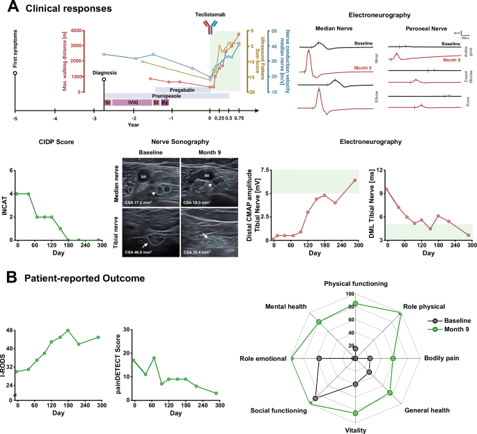 The alternative text for this image may have been generated using AI.
A Upper Panel: Left: Overview of disease course until 9 months (287 days) post-treatment follow up, prior treatment approaches, ultrasound pattern sum score (UPSS; both sides; range 0 [normal] to 44 [most severe]), and nerve conduction velocity recordings [right median nerve; normal value: > 50 m/s]. Green area: Results after teclistamab treatment. Right: Electroneurographic recordings of right median and left peroneal nerve prior treatment with teclistamab (baseline; black) and 9 months after first teclistamab dose (red) showing increased amplitudes and reduced distal motor latency (DML). Lower Panel: Left: Inflammatory Neuropathy Cause and Treatment disability score (INCAT; range 0 [normal] to 10 [most severe]) illustrating improvement of neuropathy symptoms. Nerve sonography: Median (arrow-head) and tibial nerve (arrow) sonography at baseline and month 9 showing a reduction of nerve swelling. Right: Distal compound muscle action potential (CMAP) amplitude of the right and DML recordings of the left tibial nerve showing a profound improvement. Green area: Normal values. B Patient-reported Outcome: Inflammatory Rasch-built Overall Disability Scale (I-RODS) is a patient-reported outcome scale specifically designed to assess activity and social participation limitations in patients with inflammatory polyneuropathies (range 0 [most severe] to 48 [normal]). painDETECT score: painDETECT is a patient-completed screening questionnaire designed to identify neuropathic pain components in individuals with chronic pain conditions (range 0 [no pain] to 35 [most severe]). The Short Form 36 Health Survey (SF-36): SF-36 at baseline and month 9. The SF-36 is a widely used, patient-reported questionnaire designed to measure health-related quality of life across a broad range of diseases and populations. Abbreviations: St: high-dose steroids, IVIG: intravenous immunoglobulins, Px: plasma exchange, CIDP: Chronic inflammatory demyelinating polyneuropathy, BA: brachial artery, CSA: cross-sectional area CMAP: compound muscle action potential, DML: distal motor latency. Source data are provided as a Source Data file.
Fig. 2: Patient 1: Immunological changes and serologic responses in refractory IgM-paraproteinemic autoimmune neuropathy treated with teclistamab.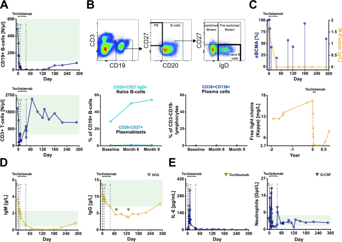 The alternative text for this image may have been generated using AI.
A Course of absolute CD19 + B-cell and CD3 + T-cell counts illustrating depletion and repopulation of CD19 + B-cells following teclistamab therapy. Green area: Normal values. B Fluorescence-Activated Cell Sorting Analysis of naïve B-cells, plasmablasts and plasma cells before and after teclistamab treatment using peripheral blood. Upper panel: excerpt of the gating strategy for naïve B-cells and plasmablasts. Lower Panel: course of naïve B-cells, plasmablasts and plasma cells (time points: baseline/d0 prior teclistamab, month 6/d179 and month 9/d287). FACS analysis was performed once at the respective time point. C Relative sBCMA levels showed a temporary decrease during the treatment phase whereas the M-protein remained undetectable until last follow-up (day 287). Free light chain (Kappa) levels were below the lower detection limit (3.7 mg/L) and increased again after approximately 7 months (day 217). sBCMA measurements were performed three times at each time point. D IgM and IgG were reduced after teclistamab treatment with subsequent increase. Purple triangles indicate time points of IVIG infusion. Green area: Normal values. E Interleukin-6 (IL-6) and neutrophil levels. Cytokine release syndrome was treated with tocilizumab (yellow triangle), neutropenia with granulocyte colony stimulating factor (G-CSF) (red triangle). Dashed lines in A, D, and E indicate time points of teclistamab injections (0.06: 0.06 mg/kg body weight, 0.3: 0.3 mg/kg body weight; 1.5: 1.5 mg/kg body weight). Source data are provided as a Source Data file.
Patient 2
A 73-year-old woman with a 13-year history of progressive sensory-ataxic gait and distal-predominant sensorimotor impairment was diagnosed with CIPNM associated with IgM-kappa paraproteins. After initial improvement of sensory deficits on high-dose steroid therapy, her disease course was progressive over the years despite multiple treatments including steroids, IVIG, plasma exchange, and rituximab, resulting in ataxic gait and distal pareses of the lower extremities with recurrent falls and fractures (Fig. 3A; Supplementary Table 1). Nerve conduction studies showed loss of motor and sensory responses in leg nerves, with profound conduction slowing of the arm nerves, and multifocal nerve swelling on ultrasound. MAG-Abs were detected, establishing the diagnosis of CIPNM-MAGAb.
Fig. 3: Patient 2: Clinical responses and patient-reported outcome in refractory MAG neuropathy treated with teclistamab.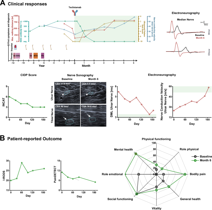 The alternative text for this image may have been generated using AI.
A Upper Panel: Left: Overview of disease course until follow-up 6 months (186 days) post-treatment, prior treatment approaches, sequelae of treatment or neuropathy, ultrasound pattern sum score (UPSS; right side; range 0 [normal] to 22 [most severe]), nerve conduction velocity recordings [left ulnar nerve; normal value: > 50 m/s], and MAG-Ab titers. Green area: Results after teclistamab treatment (note, change of x-axis intervals from years to months after teclistamab treatment). Right: Electroneurographic recordings of left median nerve prior treatment with teclistamab (baseline; black) and 6 months after first teclistamab dose (red) showing increased amplitudes and improved distal motor latency (DML). Lower Panel: Left: Inflammatory Neuropathy Cause and Treatment disability score (INCAT; range 0 [normal] to 10 [most severe]) illustrating improvement of neuropathy symptoms. Nerve sonography: Ulnar (arrow-head) and tibial nerve (arrow) sonography at baseline and month 6 showing a reduction of nerve swelling. Right: DML and nerve conduction velocity recordings of the left ulnar nerve showing a profound improvement. Green area: Normal values. B Patient-reported Outcome: Repetitive I-RODS (range 0 [most severe] to 48 [normal]) and painDETECT (range 0 [no pain] to 35 [most severe]) scores before and after teclistamab therapy. SF-36 at baseline and month 6 measuring health-related quality of life. Abbreviations: St: high-dose steroids, IVIG: intravenous immunoglobulins, RTX: rituximab, Px: plasma exchange, CIDP: Chronic inflammatory demyelinating polyneuropathy, CSA: cross-sectional area, CMAP: compound muscle action potential, DML: distal motor latency. Source data are provided as a Source Data file.
Teclistamab treatment increased walking distance over sixfold within 6 months and improved clinical scores (i.e., grip strength, I-RODS, MRC sum, INCAT, Timed Up and Go test) and nerve conduction studies (Fig. 3A, SFig. 4A-B, Supplementay Table 3). Ultrasound confirmed a marked reduction of nerve swelling (Fig. 3A, SFig. 4C). The patient also reported improvement in quality of life as well as reduced neuropathy impairment and pain (Fig. 3B). Teclistamab induced intermittent lymphopenia with a transient drop of CD3 + T-cells and complete depletion of B-cells (Fig. 4A). Again, sBCMA levels declined rapidly after treatment, followed by an increase three months after teclistamab. Remarkably, similar to IgM-kappa paraprotein levels in patient 1, M-Protein was not detectable until last follow up (Fig. 4B). Serum Nfl levels were also decreased (Fig. 4C). MAG-Ab titers decreased within the first month of therapy and remained negative until last follow up at 6 months (day 186) (Fig. 4D). Serological analysis showed a reduction of total IgM and IgG levels (Fig. 4E). Except for tetanus IgG, vaccine titers remained largely intact (SFig. 3). Following the first and second administration, the patient experienced grade 1 CRS with moderate IL-6 increase. In contrast to patient 1, no neutropenia was observed (Fig. 4F). As expected, she developed hypogammaglobulinemia, for which she received IVIG infusions (Fig. 4E). At month 5, the patient developed mild bronchitis, accompanied by moderate increases in IL-6 and neutrophil levels (Fig. 4F), without the need for antibiotic therapy or hospitalization. Clinical and paraclinical improvement remained stable with no signs of relapse until the last follow-up (month 6).
Fig. 4: Patient 2: Immunological changes and serologic responses in MAG neuropathy treated with teclistamab.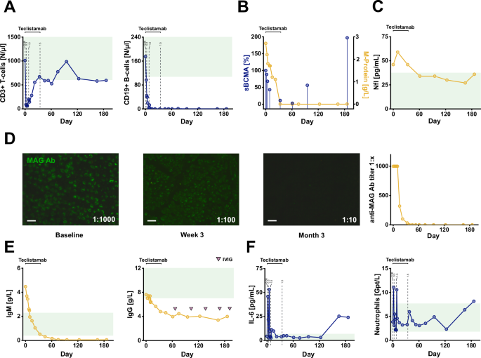 The alternative text for this image may have been generated using AI.
A CD3 + T-cell counts and CD19 + B-cell counts after teclistamab therapy. Green areas: Normal values. B Relative sBCMA levels showed a temporary decrease during the treatment phase whereas the M-protein remained undetectable until month 6. sBCMA measurements were performed three times at each time point. C Course of serum neurofilament (Nfl) levels showing reduction after teclistamab treatment. Green area: Normal values. D Left: Immunostaining of anti-MAG antibodies at baseline, 3 weeks and 3 months after first teclistamab dose showing decrease of MAG antibody titers already at week 3. MAG antibodies were negative at month 3. Right: Course of serum MAG antibody titers. Scale bars: 10 µm. E IgM and IgG levels after teclistamab treatment. Purple triangles indicate time points of supplementary IVIG infusion. Green area: Normal values. F Interleukin-6 (IL-6) levels and neutrophil levels after teclistamab. A second increase of IL-6 was observed during month 5 when the patient had a mild bronchitis. Green area: Normal values. Dashed lines in A and F indicate time points of teclistamab injections (0.06: 0.06 mg/kg body weight, 0.3: 0.3 mg/kg body weight; 1.5: 1.5 mg/kg body weight). Abbreviations: MAG-Ab: Anti-MAG antibodies, IVIG: intravenous immunoglobulin. Source data are provided as a Source Data file.