
Dual agonism of glucagon-like peptide-1 (GLP-1) and glucose-dependent insulinotropic polypeptide (GIP) receptors (GLP-1R and GIPR, respectively), in the form of once-weekly injectable tirzepatide, is highly effective for treating people living with obesity and type 2 diabetes, resulting in greater weight loss than GLP-1R agonism alone2. However, GIP biology is multimodal, and both GIPR agonism and antagonism provoke weight loss when administered together with GLP-1R agonists3. The results may be reconcilable by the existence of distinct Gipr-expressing target cell populations with different physiological roles and responsiveness. GIPR agonists decrease food intake and body weight in mice through a pathway involving Gipr-expressing GABAergic neurones, a population of which has been located in the area postrema (AP) of the brainstem4,5,6. Chemogenetic activation of Gipr-expressing neurones in either the hypothalamus or hindbrain decreases food intake in mice, suggesting more than one centrally located population of GIPR neurons regulate appetite7,8. Contrasting with these actions of GIPR activation to reduce food intake, Gipr knockout (KO) mice, and humans with impaired function GIPR variants, are protected against obesity, also probably through a central effect, as similar observations were made in global KO mice or mice lacking Gipr in nestin, synapsin or VGAT-expressing cells4,5,9,10,11,12. GIP is also anti-emetic, reducing nausea-related behaviours triggered by a variety of agents, which has been attributed to Gipr-expressing cells in the brainstem/AP13,14,15. By contrast, knockout of Gipr in GABAergic neurones did not prevent the ability of GIPR antagonists to reduce food intake6.
This study aimed to compare the physiological roles of GIPR in the AP and hypothalamus by targeted regional KO of Gipr, using a floxed Gipr mouse model (Giprfx/fx) stereotactically injected with adeno-associated virus (AAV) encoding Cre-recombinase to generate mice with KO of Gipr in either the AP (GiprAP-KO) or hypothalamus (Giprhypo-KO). Control mice were injected with AAV encoding GFP (GiprAP-GFP, Giprhypo-GFP). Whereas the AP lies outside the blood–brain barrier and has a high density of Gipr-expressing cells, most of which are GABAergic, many Gipr-expressing neurones in the hypothalamus have their cell bodies behind the blood–brain barrier and include both glutamatergic and GABAergic neurons16.
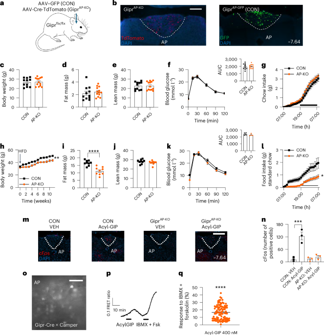
Correct targeting of the AP in GiprAP-GFP and GiprAP-KO mice (Fig. 1a) was confirmed postmortem by immunofluorescent visualisation of TdTomato, which was encoded in the AAV-Cre virus, and GFP (Fig. 1b). GiprAP-KO mice on chow diet had comparable body weight, fat mass and lean mass to control GiprAP-GFP animals (Fig. 1c–e), mirroring the previously reported phenotypes of germline global Gipr KO mice and Giprfx/fx mice crossed with Nestin-Cre to drive KO throughout the central nervous system (CNS)4,9. Chow-fed mice with KO of Gipr in GABAergic neurones achieved by crossing Giprfx/fx with VGAT-Cre strains, were also previously reported to have normal body weight, although with subtle differences in fat mass and respiratory exchange ratio (RER), and improved glucose metabolism5,11. GiprAP-KO mice had no alterations in circulating GIP, GLP-1 or insulin (Extended Data Fig. 1a–c), intraperitoneal (i.p.) glucose tolerance (Fig. 1f, with area under the curve, AUC), food intake on refeeding after the i.p. glucose tolerance test (IPGTT) or plasma glucose during ad libitum (AL) feeding (Extended Data Fig. 1e). They also showed no differences in food intake (Fig. 1g) (compared with GiprAP-GFP mice), RER, energy expenditure or ambulatory activity, as measured in metabolic cages (Extended Data Fig. 1g,h).
Fig. 1: The effects of GiprAP-KO on standard chow-fed (lean) and 45% HFD fed (DIO) mice.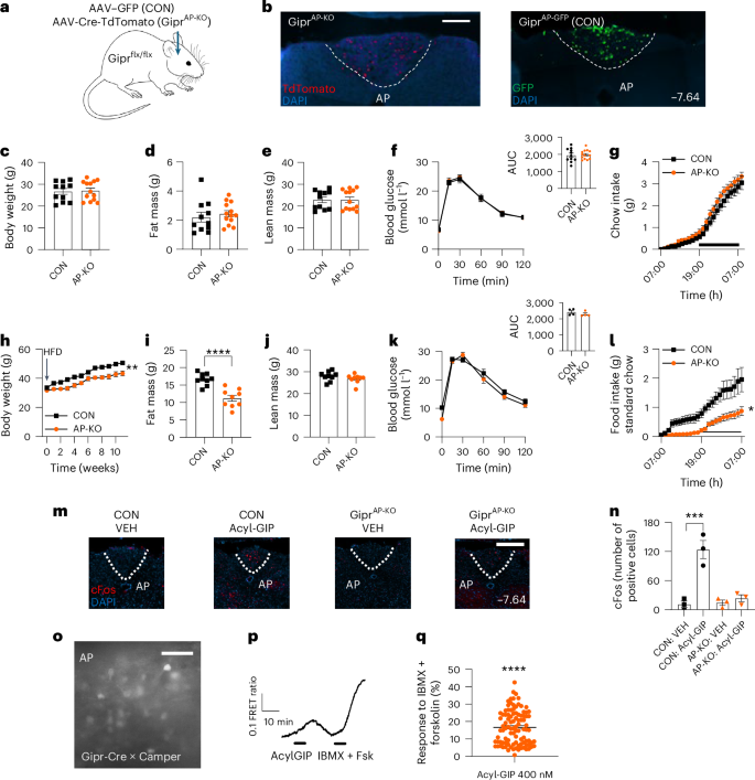
a, Schematic showing the targeting of AAV–GFP (CON, GiprAP-GFP) or AAV-Cre-TdTomato (GiprAP-KO) to the AP of Giprfx/fx mice. b, Immunohistochemistry confirming targeting of AAV-Cre (example of n = 10) and AAV–GFP (CON) (example of n = 10) (scale bar, 100 µm). Additional examples are shown in Supplementary Fig. 1. c–g, Metabolic parameters of lean mice on standard chow: body weight (c), fat mass (d), lean mass (e), IPGTT with AUC (f) and food intake in metabolic cages (g) (CON n = 11, GiprAP-KO n = 13 per group). h–l, Metabolic parameters of DIO mice on 45% HFD: body weight (h), fat mass (i), lean mass (j) (n = 9 per group for h–i), IPGTT with AUC (n = 4 per group) (k) and HFD intake in metabolic cages (l) (DIO male mice, CON n = 5, GiprAP-KO n = 4). m,n, Representative cFos images of the AP of CON and GiprAP-KO animals receiving VEH or acyl-GIP (100 nmol kg−1; scale bar, 100 µm) (m) and accompanying quantification (n) (n = 3 mice per group; additional examples are shown in Supplementary Fig. 2). o–q, cAMP imaging in ex vivo brain slices: representative image of fluorescence of the AP from Gipr-Cre × CAMPER mice (scale bar, 30 µm) (o), representative trace of cAMP-level changes monitored as FRET ratio YFP/CFP, on perfusion with acyl-GIP and IBMX-forskolin, as indicated by the bars (p) and percentage FRET response to acyl-GIP relative to IBMX-forskolin treatment (n = 101 cells from four animals monitored in nine slices) (q). Data were analysed by: two-tailed Student’s (unpaired) t-test (c–f and i–k), Welch’s test on final weight (h), Mann–Whitney test on final cumulative intake (i), one-way ANOVA with Tukey’s multiple comparisons test (n) and two-tailed one-sample t-test (q). Data are mean ± s.e.m.; *P < 0.05, **P < 0.01, ***P < 0.001, ****P < 0.0001. CON, control; DAPI, 4,6-diamidino-2-phenylindole.
GiprAP-KO mice displayed attenuated body weight gain when fed on a 45% high-fat diet (HFD), associated with reduced fat mass but preservation of lean mass (Fig. 1h–j). Fasting GIP, GLP-1 and insulin were comparable in GiprAP-KO animals and controls (Extended Data Fig. 1i–k). Glucose homeostasis as assessed by IPGTT was similar (Fig. 1k, with AUC) as was food intake following the IPGTT and AL-fed blood glucose (Extended Data Fig. 1l,m). In metabolic cages, GiprAP-KO mice showed reduced intake of HFD (Fig. 1l) and correspondingly lower RER but similar energy expenditure and ambulatory activity (Extended Data Fig. 1n–p). These characteristics are similar to those reported for GABAergic Gipr KO mice, which also had attenuated weight gain on HFD exposure, with reduced fat and lean mass5,11. Taken together, these results suggest that at least a proportion of the protection against diet-induced obesity (DIO) observed in global Gipr KO mice is attributable to loss of the population of Gipr-expressing GABAergic neurones located in the AP.
Consistent with the previously demonstrated ability of fluorescently-labelled GIP to access and activate the AP5,7, we found that acyl-GIP increased the total number of cells in the AP staining positive for cFos (a marker of neuronal activation) in control mice. Knockout of Gipr in the AP largely abolished this effect of acyl-GIP on AP cFos labelling (Fig. 1m,n). We also performed real-time imaging of cAMP levels in ex vivo hindbrain slices from Gipr-Cre × CAMPER reporter mice (Fig. 1o,p). Treatment with acyl-GIP decreased the fluorescence energy transfer ratio (FRET; YFP/CFP) in Gipr-labelled cells in the AP, reflecting an increase in cAMP levels (Fig. 1q). The response was 17 ± 1% (mean ± s.e.m., n = 101) of the maximum response to the positive control, 3-isobutyl-1-methylxanthine (IBMX) plus forskolin (Fig. 1q). These data provide high-resolution validation that acute GIP exposure directly activates (not inactivates) AP neurons.
Acyl-GIP improved i.p. glucose tolerance in wild type (WT) chow-fed mice (Extended Data Fig. 2a,b) and reduced subsequent food consumption (Extended Data Fig. 2c), consistent with previous observations4,5,17. It also reduced food intake in animals that fasted overnight and in AL-fed mice at the onset of the dark phase (Extended Data Fig. 2d,e). In a treatment-naive cohort studied in metabolic cages, administration of acyl-GIP at the onset of the dark phase decreased food intake and RER (Extended Data Fig. 2f,g) and significantly reduced body weight (Extended Data Fig. 2j). No effects were observed on energy expenditure or ambulatory activity (Extended Data Fig. 2h,i).
While previous studies have shown that CNS GABAergic neurons are necessary for the food intake- and body weight-reducing effects of GIPR agonists5,11, the anatomic region that underlies these effects remains unknown. KO of Gipr in the AP (GiprAP-KO) abolished the ability of acyl-GIP to reduce food intake at the onset of the dark phase in AL-fed animals, using GiprAP-GFP mice as a control group (Fig. 2a). Similar results were apparent in a fast-refeed paradigm—treatment with acyl-GIP significantly reduced food intake in control GiprAP-GFP animals highly motivated to feed after a 16-h fast, while this effect was lost in GiprAP-KO animals (Fig. 2b). The effects of acyl-GIP on i.p. glucose tolerance were maintained in GiprAP-KO mice as expected, consistent with direct effects of acyl-GIP on pancreatic β-cells rather than an indirect effect mediated via the AP (Extended Data Fig. 3a,b). In metabolic cages, acyl-GIP reduced food intake across the 12-h dark period, associated with a significant reduction in the RER in control GiprAP-GFP but not GiprAP-KO mice (Fig. 2c,d). No differences were observed in energy expenditure or activity (Extended Data Fig. 3c,d).
Fig. 2: Effects of acyl-GIP on food intake in lean and DIO GiprAP-KO mice.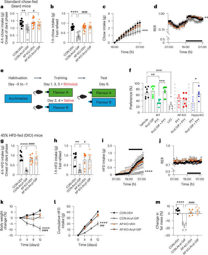
a–d, Metabolic parameters of chow-fed lean mice: food intake at the onset of the dark phase (a) and in a fast-refeed paradigm (b) of CON (n = 11, GiprAP-GFP) or GiprAP-KO (n = 13) animals receiving VEH or acyl-GIP; food intake (c) and RER (d) recorded in metabolic cages of CON (n = 7, GiprAP-GFP) and GiprAP-KO (n = 8) animals receiving VEH or acyl-GIP (in c and d, *** P < 0.001 and **P < 0.01 comparing acyl-GIP versus VEH in CON; symbols as defined in a and b). e,f, Schematic of conditioned taste preference protocol (e) and observed preference for flavour (f) (VEH = saline, acyl-GIP and PYY at 100 nmol kg−1 alone or in combination or 0.2 M LiCl) in WT, GiprAP-KO or Giprhypo-KO mice (n = 7 per group with exception of WT/acyl-GIP + PYY (n = 8) and Hypo-KO (PYY n = 6, acyl-GIP + PYY n = 5). g,h, HFD intake at the onset of the dark phase (g) and in the fast-refeed paradigm (h) of CON (n = 7) or GiprAP-KO (n = 8) DIO animals receiving VEH or acyl-GIP. i,j, HFD intake (i) and RER (j) of CON or GiprAP-KO DIO animals recorded in metabolic cages, receiving VEH (n = 6 per group) or acyl-GIP (n = 4 per group; symbols as defined in g and h). k–m, Body-weight change (n = 6 per group, with the exception of CON/acyl-GIP (n = 8)) (k), food intake (n = 5 per group) (l) and fat mass (n = 5 per group) (m) of DIO CON or GiprAP-KO animals receiving daily VEH or acyl-GIP. ****P < 0.0001 acyl-GIP versus VEH in CON; ###P < 0.001, ####P < 0.0001 acyl-GIP in CON versus GiprAP-KO; symbols as defined in l and m. Data were analysed by: one-way ANOVA with Tukey’s multiple comparisons test (a, b, g, h and m) or Sidak’s multiple comparisons test (f); two-way ANOVA including time-series data (d and j), two-way ANOVA on final food intake or body weight with uncorrected Fisher’s least significant difference (LSD) (c, i, k and l). Data are mean ± s.e.m.; *P < 0.05, **P < 0.01, ***P < 0.001 and ****P < 0.0001.
Brainstem Gipr-expressing cells have been implicated in suppressing nausea-related behaviours, such as conditioned taste avoidance (CTA), as long-acting GIPR agonists reduced CTA triggered by PYY(3-36), GLP-1 or GDF15 administration13,14,15,18,19. To determine whether GIPR in the AP is responsible for attenuating the avoidance effect of PYY, we applied a CTA assay in which exposure to PYY was paired with a novel tastant alone or in combination with acyl-GIP. In this experimental design, reduced consumption of the paired stimulus is indicative of an avoidance response (Fig. 2e). In line with previous reports, conditioning with PYY, but not acyl-GIP or vehicle (VEH) control, decreased preference for the paired taste in lean mice. Acyl-GIP abolished the avoidance effect of PYY in control GIPRAP-GFP animals but not in GIPRAP-KO animals (Fig. 2f), confirming the importance of AP Gipr-expressing cells for mediating this action of GIPR agonism. By contrast, knockout of Gipr in the hypothalamus (generated as shown in Fig. 3f) did not prevent this anti-aversive effect of acyl-GIP.
GiprAP-KO and GiprAP-GFP mice were next fed an HFD to induce DIO. At the onset of the dark phase, acyl-GIP significantly reduced food intake of DIO GiprAP-GFP but not GiprAP-KO animals (Fig. 2g), and similar results were apparent in the 16-h fast-refeed paradigm (Fig. 2h). The effects of acyl-GIP on i.p. glucose tolerance were maintained in both groups (Extended Data Fig. 3e,f). In metabolic cages, acyl-GIP reduced food intake and RER across the 12-h dark period in DIO GiprAP-GFP but not GiprAP-KO mice (Fig. 2i,j), with no differences in energy expenditure or activity (Extended Data Fig. 3g,h). Chronic daily treatment with acyl-GIP reduced body weight and food intake in control DIO GiprAP-GFP mice, associated with a significant reduction in fat mass. These effects were absent in GiprAP-KO animals treated with acyl-GIP (Fig. 2k–m). The results indicate that Gipr-expressing cells in the AP underlie the ability of GIPR agonists to reduce food intake in lean and DIO mice and, taken together with the previous report that GABAergic Gipr KO mice do not exhibit reduced food intake in response to acyl-GIP, suggest that subpopulations of GABAergic Gipr-expressing neurones located in the AP mediate both the appetite-suppressing and anti-aversive effects of exogenous GIPR agonists6.
There is emerging evidence that GIPR agonism and antagonism exert their paradoxically similar effects on weight loss via distinct neuronal populations6,12. We sought to further delineate these divergent regulatory networks by investigating whether AP GIPRs modulate responses to either GLP-1R agonism or the combination of GLP-1R agonism with GIPR antagonism. In a first study, DIO GiprAP-GFP and GiprAP-KO mice received daily injections of the GLP-1R agonist liraglutide (LIRA; 100 nmol kg−1). Over 12 days of treatment, LIRA reduced body weight, food intake and fat mass, similarly in GiprAP-KO and control GiprAP-GFP mice, compared with VEH control-injected groups (Fig. 3a–c). This indicated that the lack of Gipr in the AP did not in itself sensitize to GLP-1R agonism. In a subsequent study, lean GiprAP-KO and GiprAP-GFP mice were switched to an HFD on the same day as initiating 10 days of daily injections with either LIRA (or VEH) alone, or LIRA (or VEH) plus GIPR antagonist peptide (GIPR-antag)6. LIRA + GIPR-antag induced additional weight loss and food intake suppression compared with LIRA alone; however, there was no difference between GiprAP-KO and control GiprAP-GFP groups (Fig. 3d,e). Thus, although GIPR in the AP is required for the weight loss effects of GIPR agonists, it is not the target of therapeutic GIPR antagonists, supporting recent findings that Gipr KO in GABAergic neurons also abolished responses to GIPR agonism but not antagonism6. Nevertheless, the target for GIPR antagonists is believed to be a neuronal population in the brain, as a peptide–antibody conjugate that blocks GIPR while activating GLP-1R required central GIPR for maximal weight loss in DIO mice, as determined using mice lacking Gipr in cells expressing synapsin12.
Fig. 3: Effects of GLP-1R agonism and GIPR antagonism on GiprAP-KO and GiprHypo-KO mice.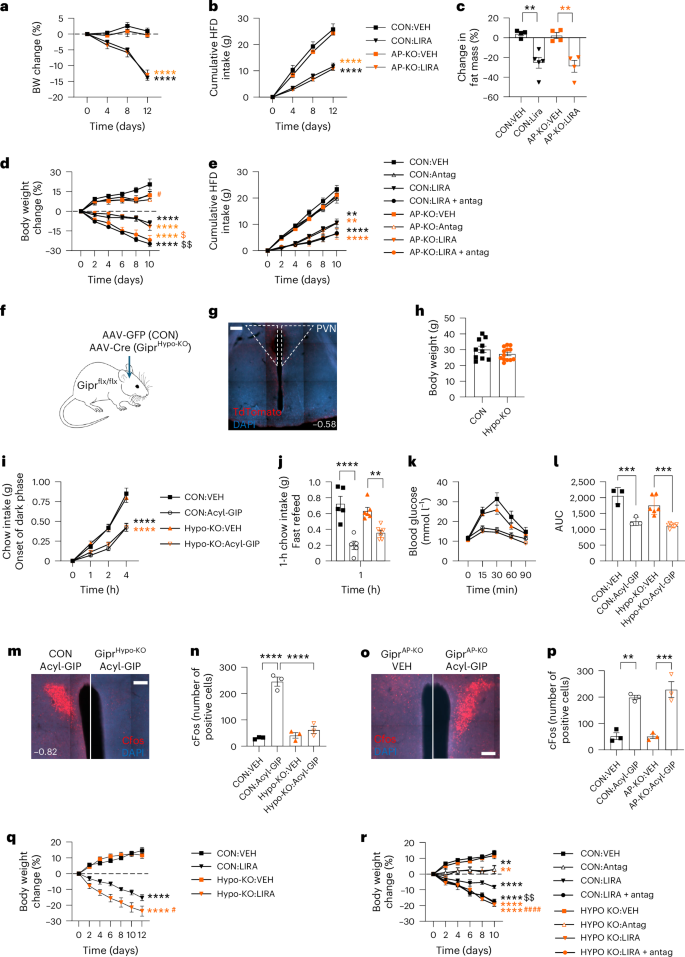
a–c, GLP-1RA responsiveness of DIO GiprAP-KO mice: body weight change (a), cumulative food intake (b) and delta fat mass (c) of CON (GiprAP-GFP) and GiprAP-KO mice receiving VEH or LIRA daily (100 nmol kg−1 (n = 4, except CON LIRA n = 5 per group; symbols as defined in b and c); black, **P < 0.01, ****P < 0.0001 LIRA versus VEH in CON; orange, **P < 0.01, ****P < 0.0001 LIRA versus VEH in GIPRAP-KO. d,e, Body weight change (d) and cumulative HFD intake (e) of CON and GiprAP-KO mice transferred to a 45% HFD and treated as indicated (CON VEH n = 5, GiprAP-KO VEH n = 5, CON antag n = 6, GiprAP-KO antag n = 6, CON LIRA n = 5, GiprAP-KO LIRA n = 5, CON LIRA + antag n = 5 and GiprAP-KO n = 7; symbols as defined in e); black, **P < 0.01, ****P < 0.0001 treatment versus VEH in CON; orange, **P < 0.01, ****P < 0.0001 treatment versus VEH in GiprAP-KO; black, $$P < 0.01 versus CON LIRA; orange, $P < 0.05 versus AP-KO LIRA; orange, #P < 0.05 antag in CON versus AP-KO. f–q, Phenotype of Giprhypo-KO mice. Schematic showing the targeting of AAV–GFP (CON) or AAV-Cre (Giprhypo-KO) to the hypothalamus of Giprfx/fx mice (f). Immunohistochemistry confirming targeting of AAV-Cre (which co-expresses TdTomato, example of n = 10, with additional examples shown in Supplementary Fig. 3; n = 10 for AAV-Cre and n = 8 (two missing owing to brain processing issues) for AAV–GFP control) (g). Body weight (CON n = 10, Giprhypo-KO n = 13) (h), chow intake at the onset of the dark phase (CON n = 10, Giprhypo-KO n = 13; black ****P < 0.0001 versus VEH in CON; orange, ****P < 0.0001 versus VEH in Giprhypo-KO) (i), chow intake in the fast-refeed paradigm of CON or Giprhypo-KO mice receiving VEH or acyl-GIP (100 nmol kg−1, CON VEH/acyl-GIP n = 5, Giprhypo-KO VEH n = 6, Giprhypo-KO acyl-GIP n = 7; symbols as defined in i) (j). IPGTT (k) with AUC of CON and Giprhypo-KO mice receiving VEH or acyl-GIP (CON VEH n = 3, CON acyl-GIP n = 3, Giprhypo-KO VEH n = 6, Giprhypo-KO acyl-GIP n = 7) (l). cFos in the PVN of CON and Giprhypo-KO animals receiving acyl-GIP (m), which is quantified (n) (n = 3 animals per group; additional examples are shown in Supplementary Fig. 4). cFos in the PVN of GiprAP-KO mice treated with VEH or acyl-GIP (o), which is quantified (p) (n = 3 animals per group; additional examples are shown in Supplementary Fig. 4). Body weight change of CON and GiprHypo-KO mice transferred to a 45% HFD and treated with VEH or LIRA as indicated (n = 6 per group; black, ****P < 0.0001 versus VEH in CON; orange, ****P < 0.0001 versus VEH in Giprhypo-KO; orange, #P < 0.05 LIRA in CON versus Giprhypo-KO) (q). r, The same as in q with the addition of GIPR-antag (co)treatment (CON VEH n = 5, Giprhypo-KO n = 6, CON antag n = 5, Giprhypo-KO antag n = 6, CON LIRA n = 7, Giprhypo-KO LIRA n = 7, CON LIRA + antag n = 6, Giprhypo-KO LIRA + antag n = 7; black, **P < 0.01, ****P < 0.0001 versus VEH in CON; orange, **P < 0.01, ****P < 0.0001 versus VEH in Giprhypo-KO; ####P < 0.0001 LIRA in CON versus Giprhypo-KO; $$LIRA versus LIRA + antag in CON). Data were analysed by: two-way ANOVA on final food intake or body weight with uncorrected Fisher’s LSD (a, b, d, e, i, qand r); one-way ANOVA with Tukey’s multiple comparisons test (c); and one-way ANOVA with Dunnett’s test for multiple comparisons (h, j, l, n and p). Data are mean ± s.e.m.; *P < 0.05, **P < 0.01, ***P < 0.001 and ****P < 0.0001.
The results mirror a previous observation from our group, that hypothalamic Gipr KO appeared to sensitize DIO mice to weight loss induced by a long-acting GLP-1R agonist—a finding that had not been the focus of that study and remained unexplained at the time but has since also been observed in global GIPR-KO mice7,11. We therefore generated new cohorts of mice lacking Gipr in the hypothalamus (Giprhypo-KO), by targeted injection of AAV-Cre or control AAV–GFP (Giprhypo-GFP) into the paraventricular nucleus (PVN) (Fig. 3f). Targeting was confirmed by the localised expression of TdTomato from the AAV-Cre virus (Fig. 3g). Giprhypo-KO mice displayed normal body weight on a chow diet (Fig. 3h). The ability of acyl-GIP to reduce food intake at the onset of the dark phase and in the 16-h fast-refeed paradigm was maintained in Giprhypo-KO mice (Fig. 3i,j), as was the effect of acyl-GIP on glucose tolerance (Fig. 3k,l). Treatment with acyl-GIP increased cFos staining in the PVN of control Giprhypo-GFP but not GIPRhypo-KO animals (Fig. 3m,n), suggesting that GIP receptors located in the hypothalamus underlie the observed hypothalamic cFos activation triggered by peripheral GIP. By contrast, KO of Gipr in the AP did not prevent cFos staining in the PVN (Fig. 3o,p).
Giprhypo-KO and control Giprhypo-GFP mice were transferred to an HFD and treated daily with either LIRA or VEH. LIRA reduced body weight in both groups, but the magnitude of the weight loss was greater in the Giprhypo-KO cohort (Fig. 3q), suggesting that these animals are sensitised to GLP-1R agonism, supporting our previous observation that sensitivity to GLP-1R agonism is enhanced in mice lacking Gipr in the hypothalamus7. We next repeated this experimental paradigm in Giprhypo-KO and Giprhypo-GFP mice, combining the initiation of HFD with a combination of LIRA (or VEH) alone or LIRA (or VEH) plus GIPR antagonist peptide (GIPR-antag). As before, LIRA caused greater weight loss in Giprhypo-KO than in Giprhypo-GFP mice, and LIRA + GIPR-antag caused more weight loss than LIRA alone in the control Giprhypo-GFP group (Fig. 3r). However, the addition of GIPR-antag to LIRA did not further enhance weight loss in the Giprhypo-KO mice, supporting the idea that the GIP receptors required for weight loss effects of GIPR antagonism are located in the hypothalamus.
Previously, treatment with a GIPR-antag was found to result in a hindbrain transcriptomic signature similar to GLP-1R agonism6. In the CNS, GLP-1 is produced by preproglucagon (Ppg) neurons with most cell bodies found in the nucleus tractus solitarius (NTS)20. We hypothesised that the reported brainstem transcriptomic changes might reflect modulation of NTS Ppg neuron activity downstream of GIPR antagonism in the hypothalamus and therefore tested the effect of GIPR-antag on mice with ablated NTS Ppg neurons (PpgNTS-Abl) generated by bilateral stereotactic injection of AAV-DTA into the NTS of Ppg-Cre or control mice (Cre-negative mice receiving AAV-DTA into the NTS) (Fig. 4a,b). Groups were treated with VEH, GIPR-antag, LIRA and LIRA + GIPR-antag. Whereas PpgNTS-Abl increased body weight gain on transfer to an HFD, and GIPR-antag attenuated this effect, ablation of the Ppg neuronal population did not attenuate the effects of any treatment arm (Fig. 4c).
Fig. 4: Interactions of GIPR antagonism with brainstem Ppg neurons and amylin receptors.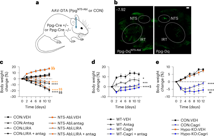
a, Schematic showing generation of Ppg-neuron ablation in the NTS by bilateral stereotactic injection of AAV-DTA into the NTS of Ppg-Cre-positive (PpgNTS-Abl) or Ppg-Cre-negative (CON) mice. b, Immunohistochemistry confirming ablation of the Ppg population in the NTS (scale bar, 100 µm, example of n = 6; additional examples shown in Supplementary Fig. 5); green fluorescence of Cre-reporters (Dq-DREADD/NuTRAP-labelling Ppg-Cre neurons) is still observed in the intermediate reticular nucleus (IRT) but not the NTS in AAV-DTA-injected animals (Ppg-DqNTS-Abl), contrasting with control Ppg-Cre animals also carrying a Dq-reporter (Ppg-Dq) shown on the right, which show GFP labelled cells in both the NTS and the IRT. c, Body weight change of CON and Ppg-DqNTS-Abl mice transferred to a 45% HFD and treated with VEH, LIRA or LIRA + GIPR-antag as indicated (CON VEH n = 6, CON antag n = 5, CON LIRA n = 6, CON LIRA + antag n = 7; PpgNTS-Abl VEH n = 5, PpgNTS-Abl antag n = 5, PpgNTS-Abl LIRA n = 6 and PpgNTS-Abl LIRA + antag n = 6); black, ****P < 0.0001 versus VEH in CON; orange, **P < 0.01, ****P < 0.0001 versus VEH in PpgNTS-Abl; orange, ##P < 0.01 VEH in CON versus PpgNTS-Abl; black, $$P < 0.01 versus LIRA in CON; orange, $$P < 0.01 versus LIRA in PpgNTS-Abl mice. d, Body weight change of WT mice transferred to a 45% HFD and treated daily with VEH, Cagri (30 nmol kg−1), GIPR-antag or Cagri + antag, as indicated (VEH n = 8, Cagri n = 8, antag n = 8, cagri + antag n = 7). *P < 0.05, ***P < 0.001, ****P < 0.0001 versus VEH; $P < 0.05 Cagri versus Cagri + antag by two-tailed unpaired t-test. e, Body weight change of CON or Giprhypo-KO mice transferred to a 45% HFD and treated daily with VEH or Cagri (30 nmol kg−1) (CON VEH n = 5, Giprhypo-KO VEH n = 5, CON Cagri n = 10, Giprhypo-KO Cagri n = 8). Data are mean ± s.e.m.; ****P < 0.0001 versus CON VEH, ##P < 0.01 Cagri in CON versus GiprRhypo-KO. Data were analysed by: two-way ANOVA on final body weight with uncorrected Fisher’s LSD (c–e).
Cagrilintide (Cagri), a long-acting amylin analogue that targets amylin receptors in the hindbrain, reduces food intake and body weight in mice21. To determine whether the weight loss sensitization effect of GIPR-antag extended to Cagri, we transferred WT mice to an HFD and treated with VEH, Cagri, GIPR-antag and the combination (Cagri + antag). Treatment with GIPR-antag reduced body weight gain, while Cagri transiently reduced body weight. The combination resulted in greater weight loss (Fig. 4d). To determine whether hypothalamic Gipr-expressing cells underlie this effect, Giprhypo-KO mice were treated with VEH or Cagri. In this cohort, Cagri induced a greater but still transient weight loss in WT animals, while in Giprhypo-KO mice, even greater weight loss was sustained to the end of the experiment (Fig. 4e).
Taken together, our results indicate that GIPR in the AP underlie the weight loss effects of GIPR agonism, whereas GIPR in the hypothalamus underlie the additional weight loss effects of adding GIPR antagonism to GLP-1R or amylin receptor agonism. Despite years of confusion around the relative benefits of GIPR agonism versus antagonism, the clinical picture that both can enhance the weight-lowering efficacy of a GLP-1R agonist seems clear. Both GIPR co-agonism (as in tirzepatide) and GIPR antagonism (as in MariTide, a bispecific hybrid molecule comprising two GLP-1R agonist peptides conjugated to a monoclonal anti-GIPR antagonistic antibody) are effective in treating people living with obesity2,3,22.
Previously, we have shown that chemogenetic activation of GIPR-expressing cells in either the hypothalamus or the hindbrain supresses food intake7,8, and GIPR-expression in oligodendrocytes has recently been linked to enhanced access of co-administered GLP-1R agonists23. We now demonstrate that GIPR agonist administration critically relies on Gipr expression in the hindbrain, while GIPR antagonism relies on hypothalamic Gipr expression—both are probably neuronal populations, given that we used hSyn-driven Cre-expression for the region-specific knockout studies. While it may be argued that our study used relatively small group sizes, the results were significant and reproducible, consistent with the large effect sizes and low variability of our experimental paradigms. The finding of distinct central populations of Gipr-expressing neurones in the AP and hypothalamus underlying responses to agonism and antagonism, respectively, goes some way towards resolving the dilemma of agonism and antagonism having similar outcomes.
Our work suggests that GIPR antagonism does not require the central production of GLP-1, as ablation of the Ppg neuron population had little effect on the weight loss outcomes. The observation that GIPR antagonism also sensitized to the non-incretin-based weight loss drug Cagri opens potential new avenues for therapeutic exploration. Future work is still, however, required to identify the neuronal networks underlying GIPR interactions in the AP and hypothalamus and their crosstalk with other appetite-regulating circuitry.